• KARM
  • Contact us
  • E-Submission
ABOUT
ARTICLE TYPES
BROWSE ARTICLES
AUTHOR INFORMATION

Articles

Original Article

The Relationship Between Tongue Pressure and Oral Dysphagia in Stroke Patients

Jong Ha Lee, MD, PhD1orcid, Hee-Sang Kim, MD, PhD1orcid, Dong Hwan Yun, MD, PhD1orcid, Jinmann Chon, MD, PhD1orcid, Yoo Jin Han, MD, PhD1orcid, Seung Don Yoo, MD, PhD2orcid, Dong Hwan Kim, MD, PhD2orcid, Seung Ah Lee, MD, PhD2orcid, Hye In Joo, MD2orcid, Ji-su Park, MS3orcid, Jin Chul Kim, MD1orcid, Yunsoo Soh, MD2orcid
Annals of Rehabilitation Medicine 2016;40(4):620-628.
Published online: August 24, 2016

1Department of Rehabilitation & Physical Medicine, Kyung-Hee Medical Center, Seoul, Korea.

2Department of Rehabilitation & Physical Medicine, Kyung-Hee University Hospital at Gangdong, Seoul, Korea.

3Department of Occupational Therapy, Inje University Busan Paik Hospital, Busan, Korea.

Corresponding author: Yunsoo Soh. Department of Rehabilitation & Physical Medicine, Kyung-Hee University Hospital at Gangdong, 892 Dongnam-ro, Gangdong-gu, Seoul 05278, Korea. Tel: +82-2-440-7246, Fax: +82-2-440-7260, soyuns@gmail.com
• Received: August 13, 2015   • Accepted: November 3, 2015

Copyright © 2016 by Korean Academy of Rehabilitation Medicine

This is an open-access article distributed under the terms of the Creative Commons Attribution Non-Commercial License (http://creativecommons.org/licenses/by-nc/4.0) which permits unrestricted noncommercial use, distribution, and reproduction in any medium, provided the original work is properly cited.

  • 9,248 Views
  • 186 Download
  • 60 Web of Science
  • 69 Crossref
  • 62 Scopus
prev next
  • Objective
    To evaluate the relationships between tongue pressure and different aspects of the oral-phase swallowing function.
  • Methods
    We included 96 stroke patients with dysphagia, ranging in age from 40 to 88 years (mean, 63.7 years). Measurements of tongue pressure were obtained with the Iowa Oral Performance Instrument, a device with established normative data. Three trials of maximum performance were performed for lip closure pressure (LP), anterior hard palate-to-tongue pressure (AP), and posterior hard palate-to-tongue pressure (PP); buccal-to-tongue pressures on both sides were also recorded (buccal-to-tongue pressure, on the weak side [BW]; buccal-to-tongue pressure, on the healthy side [BH]). The average pressure in each result was compared between the groups. Clinical evaluation of the swallowing function was performed with a videofluoroscopic swallowing study.
  • Results
    The average maximum AP and PP values in the intact LC group were significantly higher than those in the inadequate lip closure group (AP, p=0.003; PP, p<0.001). AP and PP showed significant relationships with bolus formation (BF), mastication, premature bolus loss (PBL), tongue to palate contact (TP), and oral transit time (OTT). Furthermore, LP, BW, and BH values were significantly higher in the groups with intact mastication, without PBL and intact TP.
  • Conclusion
    These findings indicate that the tongue pressure appears to be closely related to the oral-phase swallowing function in post-stroke patients, especially BF, mastication, PBL, TP and OTT.
Dysphagia is a common complication in post-stroke patients, with reported incidences varying between 29% and 67% [1]. Post-stroke dysphagia is associated with serious complications such as aspiration, a prolonged length of hospital stay, and increased mortality. Early detection and treatment of dysphagia are crucial issues in post-stroke patients [2].
The process of normal swallowing consists of a three-stage sequential model. The swallowing process is divided into the oral, pharyngeal, and esophageal stages according to the location of the bolus [3]. The movement of the tongue is an important contributor to the oral-stage swallowing. It plays a crucial role in maintaining a cohesive bolus while manipulating the bolus during mastication, and propelling the bolus out of the oral cavity and through the larynx. In stroke patients, dysfunctions of lower tongue pressure such as lingual discoordination can cause a range of problems in the oral-phase swallowing [4]. Clinically, tongue pressure has been reported to be a good predictor of the presence of oral-phase dysphagia [5678]. Until now, there have been few studies that evaluated tongue pressure in relation to different aspects of the oral-phase swallowing function in patients with post-stroke dysphagia.
Various bedside tests have been applied for evaluating dysphagia after a stroke. However, almost half of the patients continue to suffer from silent aspiration. In such cases, a videofluoroscopic swallowing study (VFSS) is the gold standard for detecting dysphagia [910]. Clinical evaluation of swallowing function was performed with a VFSS, which provided visual assessment of the entire process of deglutitive movement and bolus transport [11].
In addition to the oral-phase swallowing function, we also assessed the relationship between tongue pressure and the patients' functional outcomes. Previous studies have found that patients with dysphagia had significantly lower cognitive function as well as functional evaluation score [12]. In this study, the Korean version of Mini Mental State Examination (K-MMSE) and the Korean version of Modified Barthel Index (K-MBI) scores were used to evaluate cognitive function and activities of daily living (ADL), respectively.
The aims of this study were (1) to evaluate the relationships between tongue pressure and different aspects of the oral-phase swallowing function and (2) to evaluate the relationships between tongue pressure and functional outcomes.
Subjects were recruited from a group of patients referred to the Department of Rehabilitation and Physical Medicine, Kyung-Hee Medical Center, Seoul, Korea for swallowing evaluation between May 2011 and January 2013. Both inpatients and outpatients were included in the study. The subjects included stroke patients (1) who had a first-ever unilateral stroke, confirmed by MRI and/or CT and were older than 50 years of age; (2) who had symptoms and/or signs of dysphagia after onset of the stroke (choking, cough, wet voice after swallowing or a history of aspiration pneumonia); (3) who were evaluated by VFSS; and (4) who could follow the physiotherapist's orders.
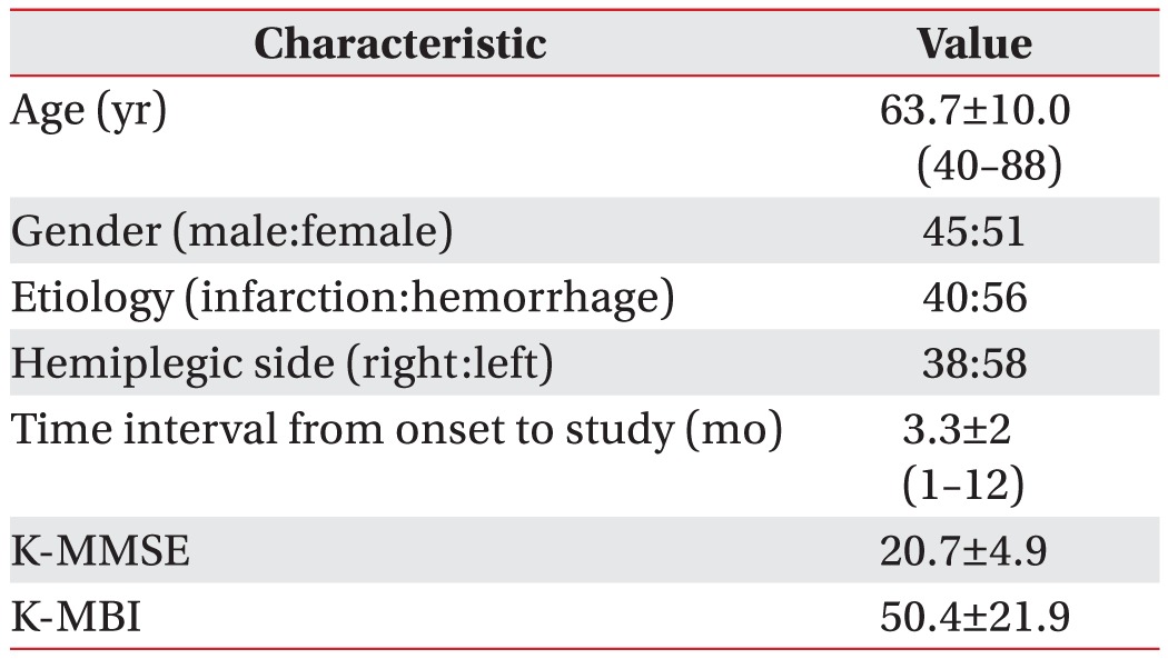
All patients were alert, had an adequate level of consciousness and were able to complete the tasks required for the study. Analysis was performed in 96 patients with non-traumatic cerebral infarction or hemorrhage. The patients ranged in age from 40 to 88 years, with a mean age of 63.75 years. The mean age of the 45 male participants was 63.57 years (range, 41–88 years), and that of the 51 female patients was 63.91 years (range, 40–82 years). Measurements were performed 1–12 months after the stroke (median, 3.8 months post-onset). The dysphagic group included 40 patients (41.6%) with cerebral infarction and 56 patients (58.3%) with cerebral hemorrhage. Of these patients, 38 (39.5%) had right hemiplegic stroke and 58 (60.4%) had left hemiplegia (Table 1).
VFSS protocol
The swallowing ability of patients with stroke was evaluated by a VFSS, which was performed as described below [513]. A modified version of Logemann [14] protocol was used. Patients were initially instructed to sit upright for 5 seconds to assess their ability to maintain posture. The videofluoroscopy instrument was positioned lateral to the patient. Liquid barium (40% weight/volume, barium sulfate, Solotop suspension) was diluted with yogurt (the viscosity was around 1,000 centipoise [cP]). First, the patients were given 5 mL of yogurt to swallow using a syringe. Afterwards, 4 g of cooked rice with barium was given using a spoon (the viscosity was above 1,750 cP). If the patients swallowed without aspiration or penetration, 5 mL of water with diluted barium (the viscosity was 1–50 cP) was given using a syringe. The entire clinical procedure was recorded with a video camera. Results of the first videofluoroscopy procedure were used for our analysis.
Interpretation
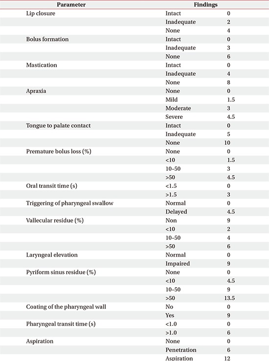
The swallowing process in the oral phase was evaluated with the videofluoroscopic dysphagia scale (VDS) (Appendix 1) [15], which consists of the following 14 items: oral phase (lip closure, bolus formation, mastication, apraxia, premature bolus loss, and oral transit time) and pharyngeal phase (pharyngeal triggering, vallecular and pyriform sinus residues, laryngeal elevation and epiglottic closure, pharyngeal coating, pharyngeal transit time, and aspiration) that can be assessed by the VFSS. All of the outcome scales were rated by two skilled physicians who had been trained in VFSS for at least 3 years. The physicians analyzed the video files and drew consensual conclusions.
In the oral phase, the completeness of lip closure (LC), bolus formation (BF), mastication, and tongue-to-palate contact (TP) were measured. The examiner categorized the results into three levels according to the degree (intact, 0; inadequate, 4; none, 8). Oral transit time (OTT) was also measured from the time the bolus started to move from the oral cavity to the lower edge of the mandibular ramus. More than 1.5 seconds of OTT was defined as the delayed oral phase. The degree of apraxia was evaluated in terms of four levels (none, 0; mild, 1.5; moderate, 3; severe, 4.5).
The amount of premature bolus loss (PBL), which pertains to the bolus drop into the pharynx from the oral cavity before the swallowing reflex, was classified into four levels (grade 0, none; grade 1, 10% of bolus; grade 2, from 10% to 50% of bolus; grade 3, >50% of bolus) [15]. To minimize the examiners' subjective error, we also compared the results by dividing the group based on the presence of PBL.
Measurement of tongue pressure
Subjective measures of tongue pressure were obtained using the Iowa Oral Performance Instrument (IOPI), a device that has been described in several previous reports [1617]. The IOPI consists of a pressure transducer and an amplifier that displays, in kilopascals (kPa), the pressure exerted on an air-filled bulb (Fig. 1).
Subjects were seated upright and asked to press their tongue against the IOPI bulb as hard as possible. Only valid (i.e., the bulb was properly positioned and pressure was applied by the tongue) trials of maximum performance for lip closure pressure (LP), anterior hard palate-to-tongue pressure (AP), posterior hard palate-to-tongue pressure (PP), and buccal pressure on both sides were recorded (buccal-to-tongue pressure on the weak side [BW]; buccal-to-tongue pressure on the healthy side [BH]). Each parameter was evaluated separately. The average pressure in the three trials was compared between the groups.
Functional outcomes
Scores on the MBI were used to evaluate ADL. The K-MBI score is commonly used as a valid and reliable measure to determine functional ability with respect to 10 domains, and total scores range from 0 (totally dependent) to 100 (totally independent) [18]. K-MMSE, which was used to evaluate cognitive function, tests five domains of cognitive function: orientation, attention, calculation, language, and construction. Total scores range from 0 to 30, and higher K-MMSE scores indicate better cognition [19].
Statistical analysis
We compared the groups statistically using the SPSS software ver. 20.0 K for Windows (SPSS Inc., Chicago, IL, USA).
The Mann–Whitney U-test was used to analyze the relationships between tongue pressure and different aspects of the oral-phase swallowing function. The Jonckheere–Terpstra test was used to compare the linear correlation between PBL and tongue pressure. Statistical significance was set at p<0.05.
The patients were divided into groups based on the intactness of the oral phase according to the VDS scale. The average differences in tongue pressure were observed between the groups. Apraxia was not compared in this study because only 1 patient had oral-phase apraxia.
Lip closure
Patients were classified into three groups (intact, 0; inadequate, 2; none, 4), but none of the patients met the criteria for the 'none' group. The intact and inadequate groups were compared. The average maximum LP was 13.8 kPa (SD=4.1) in the intact LC group (n=81) and 12.0 kPa (SD=5.3) in the inadequate LC group (n=15); this difference was not statistically significant (p=0.181). However, the average maximum AP and PP values in the intact LC group were significantly higher than those in the inadequate LC group (AP, p=0.003; PP, p<0.001). The two groups did not differ significantly with respect to K-MMSE and MBI scores (Table 2).
Bolus formation
Eighteen patients showed intact BF and 78 patients showed inadequate BF. BF was significantly correlated with AP and PP. The average maximum AP and PP values were significantly higher in the intact BF group than in the inadequate BF group (AP, p<0.001; PP, p<0.001). However, there was no significant difference between the groups with respect to the other parameters (Table 2).
Mastication
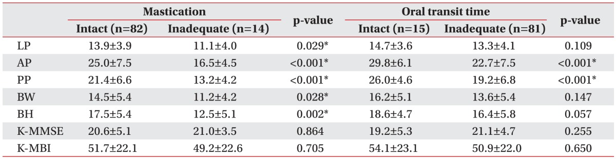
Patients with normal mastication (n=82) had significantly higher AP and PP values than patients with inadequate mastication (n=14; AP, p<0.001; PP, p<0.001). Additionally, there was a significant difference in buccal-totongue pressure on both sides and LP between the intact mastication and inadequate mastication groups (Table 3).
Oral transit time
Like other dysphagia scales, AP and PP were significantly correlated with OTT in our study. This correlation was negative, indicating that increased pressure was associated with shorter OTT. According to the VDS, patients were divided into two groups depending on OTT (<1.5 seconds vs. ≥1.5 seconds). The average maximum AP and PP values in the normal OTT group were significantly higher than those in the delayed OTT group (AP, p<0.001; PP, p<0.001). There was no significant correlation between K-MMSE scores and OTT (Table 3).
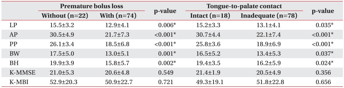
Premature bolus loss
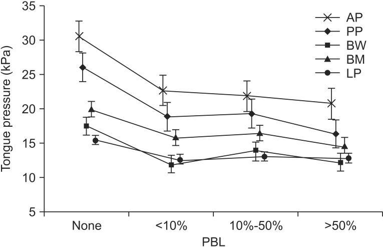
When we divided the patients into groups according to the presence of PBL, tongue pressure was associated with PBL (AP, p<0.001; PP, p<0.001; BW, p=0.001; BH, p=0.002; LP, p=0.006) (Table 4), as was the case with the other oral-phase dysphagia scales. The VDS classifies the amount of premature bolus loss into four levels. We compared these levels with tongue pressure, and the linear correlation between PBL and tongue pressure was tested with the Jonckheere–Terpstra rank correlation test. We found that a larger amount of PBL was associated with a lower maximal tongue pressure (AP, p<0.001; PP, p<0.001; BW, p=0.011; BH, p=0.008; LP, p=0.033) (Fig. 2).
Tongue-to-palate contact
Like PBL, TP was correlated with all five parameters (AP, p<0.001; PP, p<0.001; BW, p=0.037; BH, p=0.024; LP, p=0.035) (Table 4).
Previous studies have revealed that tongue pressure is a good predictor of the presence of oral-phase swallowing impairment, especially for BF, mastication, and oral clearance [820].
Robbins et al. [21] reported that an 8-week lingual resistance exercise program showed promising results with respect to preventing dysphagia due to sarcopenia and serving as a treatment strategy for patients with lingual weakness and swallowing disorders. Their results suggest that increasing tongue pressure with lingual exercises could be helpful in improving oral dysphagia.
We found that AP and PP were related to all phases of oral swallowing. This indicates that forward and backward tongue movements, such as tongue protrusion and retraction, were more important factors in oral-phase swallowing than lateral tongue movements. Only PBL, TP, and mastication were correlated with buccal-to-tongue pressure on both sides.
In the present study, LC was not related to the degree of LP, but it was significantly associated with AP and PP. In stroke patients, inadequate LC is frequently found in patients with facial palsy. Hagg and Anniko [22], who studied the association between LP and swallowing capacity, found that LP did not differ according to whether or not facial palsy was present. However, LP was significantly lower in stroke patients than in healthy subjects. These factors may indicate that LC has no relation with LP.
Tongue movement is also an important factor in holding the bolus. While drinking a liquid substance, the posterior oral cavity is sealed by tongue–palate contact during the oral preparatory stage when the bolus is held in the oral cavity [23]. Clark et al. [8] measured tongue strength with the IOPI and found that holding and manipulating the bolus were most strongly correlated with subjective measures of tongue strength. Like this previous study, we also found that PBL was related to all five parameters.
To the best of our knowledge, this is the first published study that examined the relationship between tongue pressure and functional outcomes. The K-MMSE and K-MBI results showed no significant differences in each oral phase between the intact and inadequate groups. Moon et al. [24] studied the cognition factors affecting oral-phase dysphagia in stroke patients. In contrast to our study, they found that inadequate LC and PBL were associated with low K-MMSE scores. However, we found no correlation between K-MMSE scores and any phase of oral dysphagia. Unlike the previous study, we included dementia patients who could follow our simple steps in order. Thus, their inclusion criteria differed from our criteria.
This study has several limitations. First, the evaluation of mastication, TP and amount of PBL may vary from physician to physician. Although two experienced rehabilitation physicians interpreted the VFSS, the results were subjective. Kim et al. [25] also demonstrated that VDS showed a lower rate of inter-rater agreement in the oral-phase parameters. Second, there was no healthy control group. Third, the timing of the study varied, as each patient had a different onset time depending on when the patient was referred to our rehabilitation center. Finally, the possible correlation between the location of the brain lesion and tongue pressure was not considered in this study. In cases of medullary infarction, with involvement of the hypoglossal nerve, ipsilateral tongue deviation and tongue movement disorder may be observed. Further study is needed to evaluate these limitations.
In summary, the findings of this study demonstrate the relationship of tongue weakness, as measured by tongue pressure, with oral-phase dysphagia in post-stroke patients. This suggests that patients with tongue-movement weakness during oral swallowing are at high risk for oral-phase dysphagia. However, the ways in which tongue weakness affects oral-phase swallowing remain unclear. Thus, further study is needed to precisely determine the relationship between tongue pressure and the oral-phase swallowing function.

CONFLICT OF INTEREST: No potential conflict of interest relevant to this article was reported.

  • 1. Martino R, Foley N, Bhogal S, Diamant N, Speechley M, Teasell R. Dysphagia after stroke: incidence, diagnosis, and pulmonary complications. Stroke 2005;36:2756-2763.
  • 2. Smithard DG, O'Neill PA, Parks C, Morris J. Complications and outcome after acute stroke. Does dysphagia matter? Stroke 1996;27:1200-1204.
  • 3. Dodds WJ, Stewart ET, Logemann JA. Physiology and radiology of the normal oral and pharyngeal phases of swallowing. AJR Am J Roentgenol 1990;154:953-963.
  • 4. Daniels SK, Brailey K, Foundas AL. Lingual discoordination and dysphagia following acute stroke: analyses of lesion localization. Dysphagia 1999;14:85-92.
  • 5. Logemann JA. Evaluation and treatment of swallowing disorders. 2nd ed. Austin: Pro-Ed; 1998.
  • 6. Konaka K, Kondo J, Hirota N, Tamine K, Hori K, Ono T, et al. Relationship between tongue pressure and dysphagia in stroke patients. Eur Neurol 2010;64:101-107.
  • 7. Ku DN, Ma PP, McConnel FM, Cerenko D. A kinematic study of the oropharyngeal swallowing of a liquid. Ann Biomed Eng 1990;18:655-669.
  • 8. Clark HM, Henson PA, Barber WD, Stierwalt JA, Sherrill M. Relationships among subjective and objective measures of tongue strength and oral phase swallowing impairments. Am J Speech Lang Pathol 2003;12:40-50.
  • 9. Horner J, Massey EW. Silent aspiration following stroke. Neurology 1988;38:317-319.
  • 10. Roth EJ. Medical complications encountered in stroke rehabilitation. Phys Med Rehabil Clin North Am 1991;2:563-577.
  • 11. DePippo KL, Holas MA, Reding MJ, Mandel FS, Lesser ML. Dysphagia therapy following stroke: a controlled trial. Neurology 1994;44:1655-1660.
  • 12. Falsetti P, Acciai C, Palilla R, Bosi M, Carpinteri F, Zingarelli A, et al. Oropharyngeal dysphagia after stroke: incidence, diagnosis, and clinical predictors in patients admitted to a neurorehabilitation unit. J Stroke Cerebrovasc Dis 2009;18:329-335.
  • 13. Han TR, Paik NJ, Park JW. Quantifying swallowing function after stroke: a functional dysphagia scale based on videofluoroscopic studies. Arch Phys Med Rehabil 2001;82:677-682.
  • 14. Logemann JA. Manual for the videofluorographic study of swallowing. 2nd ed. Austin: Pro-Ed; 1993.
  • 15. Han TR, Paik NJ, Park JW, Kwon BS. The prediction of persistent dysphagia beyond six months after stroke. Dysphagia 2008;23:59-64.
  • 16. Robbins J, Levine R, Wood J, Roecker EB, Luschei E. Age effects on lingual pressure generation as a risk factor for dysphagia. J Gerontol A Biol Sci Med Sci 1995;50:M257-M262.
  • 17. Park JS, You SJ, Kim JY, Yeo SG, Lee JH. Differences in orofacial muscle strength according to age and sex in East Asian healthy adults. Am J Phys Med Rehabil 2015;94:677-686.
  • 18. Shah S, Vanclay F, Cooper B. Improving the sensitivity of the Barthel Index for stroke rehabilitation. J Clin Epidemiol 1989;42:703-709.
  • 19. Kang Y, Na DL, Hahn S. A validity study on the Korean Mini-Mental State Examination (K-MMSE) in dementia patients. J Korean Neurol Assoc 1997;15:300-308.
  • 20. Pouderoux P, Kahrilas PJ. Deglutitive tongue force modulation by volition, volume, and viscosity in humans. Gastroenterology 1995;108:1418-1426.
  • 21. Robbins J, Gangnon RE, Theis SM, Kays SA, Hewitt AL, Hind JA. The effects of lingual exercise on swallowing in older adults. J Am Geriatr Soc 2005;53:1483-1489.
  • 22. Hagg M, Anniko M. Influence of lip force on swallowing capacity in stroke patients and in healthy subjects. Acta Otolaryngol 2010;130:1204-1208.
  • 23. Matsuo K, Palmer JB. Anatomy and physiology of feeding and swallowing: normal and abnormal. Phys Med Rehabil Clin N Am 2008;19:691-707.
  • 24. Moon HI, Pyun SB, Kwon HK. Correlation between location of brain lesion and cognitive function and findings of videofluoroscopic swallowing study. Ann Rehabil Med 2012;36:347-355.
  • 25. Kim DH, Choi KH, Kim HM, Koo JH, Kim BR, Kim TW, et al. Inter-rater reliability of videofluoroscopic dysphagia scale. Ann Rehabil Med 2012;36:791-796.
Appendix 1

Videofluoroscopic dysphagia scale [15]

arm-40-620-a001.jpg
Fig. 1

Positioning of the air-filled lingual pressure sensor of the Iowa Oral Performance Instrument between the tongue and an oral structure.

arm-40-620-g001.jpg
Fig. 2

Correlation between premature bolus loss (PBL) and mean tongue pressure. A larger amount of PBL was associated with a lower maximal tongue pressure (Jonckheere– Terpstra rank correlation test, p<0.05). AP, anterior hard palate-to-tongue pressure; PP, posterior hard palate-to-tongue pressure; BW, buccal-to-tongue pressure on the weak side; BH, buccal-to-tongue pressure on the healthy side; LP, lip closure pressure.

arm-40-620-g002.jpg
Table 1

Characteristics of stroke patients (n=96)

Values are presented as mean±standard deviation (range).

K-MMSE, the Korean version of Mini-Mental State Examination; K-MBI, the Korean version of Modified Barthel Index.

arm-40-620-i001.jpg
Table 2

The relationships of LP, tongue pressure, and functional outcome with lip closure and bolus formation (unit, kPa)

Values are presented as mean±standard deviation.

LP, lip pressure; AP, anterior hard palate-to-tongue pressure; PP, posterior hard palate-to-tongue pressure; BW, buccal-to-tongue pressure on the weak side; BH, buccal-to-tongue pressure on the healthy side; K-MMSE, the Korean version of Mini-Mental State Examination; K-MBI, the Korean version of Modified Barthel Index.

*p<0.05, Mann–Whitney U test.

arm-40-620-i002.jpg
Table 3

The relationships of LP, tongue pressure, and functional outcome with mastication and oral transit time (unit, kPa)

Values are presented as mean±standard deviation.

LP, lip pressure; AP, anterior hard palate-to-tongue pressure; PP, posterior hard palate-to-tongue pressure; BW, buccal-to-tongue pressure on the weak side; BH, buccal-to-tongue pressure on the healthy side; K-MMSE, the Korean version of Mini-Mental State Examination; K-MBI, the Korean version of Modified Barthel Index.

*p<0.05, Mann–Whitney U test.

arm-40-620-i003.jpg
Table 4

The relationships of LP, tongue pressure, and functional outcome with premature bolus loss and tongue-topalate contact (unit, kPa)

Values are presented as mean±standard deviation.

LP, lip pressure; AP, anterior hard palate-to-tongue pressure; PP, posterior hard palate-to-tongue pressure; BW, buccal-to-tongue pressure on the weak side; BH, buccal-to-tongue pressure on the healthy side; K-MMSE, the Korean version of Mini-Mental State Examination; K-MBI, the Korean version of Modified Barthel Index.

*p<0.05, Mann–Whitney U test.

arm-40-620-i004.jpg

Figure & Data

References

    Citations

    Citations to this article as recorded by  
    • Application of Action Observation Therapy in Swallowing Rehabilitation: A Randomised Controlled Study
      Arzucan Toksal Uçar, Bilge Piri Çınar, Banu Alıcıoğlu, Geylan Bostan, Sevil Bilgin
      Journal of Oral Rehabilitation.2025; 52(3): 320.     CrossRef
    • Đánh giá áp lực lưỡi và sức bền lưỡi của bệnh nhân đến khám tại Trung tâm lâm sàng Răng Hàm Mặt Trường Đại học Quốc tế Hồng Bàng
      Trịnh Minh Trí Trịnh Minh Trí , Văn Hồng Phượng Văn Hồng Phượng, Phạm Nguyên Quân Phạm Nguyên Quân
      Tạp Chí Khoa học Trường Đại học Quốc tế Hồng Bàng.2025; : 35.     CrossRef
    • Application of Orofacial Muscle Strength Measurement to Screen for Penetration/Aspiration Risk in Older Adults With Sarcopenia: A Diagnostic Accuracy Study
      Na‐Kyoung Hwang, Tae‐Hyung Yoon, Ji‐Su Park
      Journal of Oral Rehabilitation.2025;[Epub]     CrossRef
    • Swallowing Therapy Effects on Oral Muscle Mass, Tongue Function, Swallowing, and Nutrition in Stroke Patients with Dysphagia
      Guan-Yi Li, Yu-Chi Huang, Jia-Ying Wu, Chau-Peng Leong, Chung-Hui Cheng
      NeuroRehabilitation: An International, Interdisciplinary Journal.2025;[Epub]     CrossRef
    • Therapy of swallowing and speech problem in patients with progressive supranuclear palsy
      Mila Bunijevac
      Srpski arhiv za celokupno lekarstvo.2025; 153(1-2): 78.     CrossRef
    • Dysphagia Prevention Strategies for Community‐Dwelling Older Adults: A Scoping Review
      Kristamuliana Kristamuliana, Eri Yanuar Achmad Budi Sunaryo, Miaofen Yen, Huan‐Fang Lee
      Journal of Oral Rehabilitation.2025;[Epub]     CrossRef
    • Predictors of Swallowing-Related Quality of Life in United States Veterans with Dysphagia
      Hiba Najeeb, Kailey J. Augenstein, Joanne Yee, Aimee T. Broman, Nicole Rogus-Pulia, Ashwini Namasivayam-MacDonald
      Dysphagia.2024; 39(1): 150.     CrossRef
    • Association between tongue pressure and oral status and activities of daily living in stroke patients admitted to a convalescent rehabilitation unit
      Shizuka Ninomiya, Wataru Fujii, Erika Matsumoto, Kiichiro Yamaguchi, Masao Hiratsuka
      Clinical and Experimental Dental Research.2024;[Epub]     CrossRef
    • Oral Health-Related Factors Associated with Dysphagia Risk among Older, Healthy, Community-Dwelling Korean Adults: A Pilot Study
      Da-Som Lee, Hee-Eun Kim, Jun-Seon Choi
      Healthcare.2024; 12(2): 267.     CrossRef
    • Conceptualizing Adult Dysphagia in the United States Within the International Classification of Functioning, Disability and Health (ICF)
      Melani Zuckerman, Shawn Wang, Asako Kaneoka, Wendy J. Coster, Rebecca Leonard, Susan E. Langmore, Jessica M. Pisegna
      Archives of Physical Medicine and Rehabilitation.2024; 105(5): 1008.     CrossRef
    • Prevalence of swallowing disorder in different dementia subtypes among older adults: a meta-analysis
      Alfiani Rahmi Putri, Yu-Hao Chu, Ruey Chen, Kai-Jo Chiang, Kondwani Joseph Banda, Doresses Liu, Hui-Chen Lin, Shu-Fen Niu, Kuei-Ru Chou
      Age and Ageing.2024;[Epub]     CrossRef
    • Improvement of oral function and its impact on oral food intake in subacute stroke patients: A prospective study with dental intervention
      Yu Sekimoto, Koichiro Matsuo, Ayu Sakai, Seiko Shibata, Shunsuke Minakuchi
      Journal of Oral Rehabilitation.2024; 51(8): 1365.     CrossRef
    • Ultrasound-based visualization measurement of hyoid-mandibular motion for assessing the efficacy of acupuncture in treating post-stroke dysphagia: A clinical study
      Lu Zhang, Xiaoyang Lian, Jie Chen, Chuchu Zhang, Gang Ouyang
      Journal of Radiation Research and Applied Sciences.2024; 17(3): 100991.     CrossRef
    • Treatment of Esophageal Achalasia With Sarcopenic Dysphagia by Rehabilitation and Nutritional Support: A Case Report
      Fumiko Furukawa, Kiyohito Kakita
      Cureus.2024;[Epub]     CrossRef
    • Effect of home‐based tongue‐strengthening exercise using a portable tool on oropharyngeal muscles in older adults with sarcopenic dysphagia: A randomised controlled study
      Tae‐Hyung Yoon, Motoyoshi Morishita, Na‐Mi Han, Ji‐Su Park
      Journal of Oral Rehabilitation.2024; 51(11): 2270.     CrossRef
    • Immediate effects of simple palatal augmentation prosthesis in maximum tongue pressure and swallowing functions for acute stroke patients: An observational study
      Yuu Hashimoto, Yasunori Umemoto, Shigeru Suzuki, Yuri Miyazaki, Yukihide Nishimura, Ken Kouda
      Journal of Prosthodontic Research.2024; 69(1): 76.     CrossRef
    • Orofacial Muscle Strength and Associated Potential Factors in Healthy Korean Community-Dwelling Older Adults: A Pilot Cross-Sectional Study
      Da-Som Lee, Ji-Youn Kim, Jun-Seon Choi
      Applied Sciences.2024; 14(22): 10560.     CrossRef
    • Nutritional management for late complications of radiotherapy
      Jiahua lyu, Yue Su, Hansong Bai, Hao Kuang, Churong Li, Xiumei Zheng, Long Liang, Lu li, Diou Cheng, Tao Li
      Holistic Integrative Oncology.2024;[Epub]     CrossRef
    • Application of Ultrasonography in Neurogenic Dysphagia: A Systematic Review
      Paola Potente, Alex Buoite Stella, Monica Vidotto, Michelle Passerini, Giovanni Furlanis, Marcello Naccarato, Paolo Manganotti
      Dysphagia.2023; 38(1): 65.     CrossRef
    • Perioperative Decrease in Tongue Pressure is an Intervenable Predictor of Aspiration After Esophagectomy
      Kazuhiro Kojima, Takuya Fukushima, Daisuke Kurita, Aiko Matsuoka, Koshiro Ishiyama, Junya Oguma, Hiroyuki Daiko
      Dysphagia.2023; 38(4): 1147.     CrossRef
    • Maximum lingual pressure impacts both swallowing safety and efficiency in individuals with amyotrophic lateral sclerosis
      Raele Donetha Robison, Lauren DiBiase, Amber Anderson, James P. Wymer, Emily K. Plowman
      Neurogastroenterology & Motility.2023;[Epub]     CrossRef
    • Effects of Device-Facilitated Lingual Strengthening Therapy on Dysphagia Related Outcomes in Patients Post-Stroke: A Randomized Controlled Trial
      Brittany N. Krekeler, Joanne Yee, Atsuko Kurosu, Fauzia Osman, Rodolfo Peña-Chávez, Glen Leverson, Brittany Young, Justin Sattin, Molly Knigge, Susan Thibeault, Nicole Rogus-Pulia
      Dysphagia.2023; 38(6): 1551.     CrossRef
    • Evaluation of usefulness of tongue pressure measurement device for dysphagia associated with treatment of patients with head and neck cancer (ELEVATE)
      Akihisa Tanaka, Hirokazu Uemura, Takahiro Kimura, Ari Nishimura, Kumiko Aoki, Shintaro Otsuka, Keita Ueda, Tadashi Kitahara
      Medicine.2023; 102(26): e33954.     CrossRef
    • Correlation Between Articulatory Diadochokinetic Parameters and Dysphagia Parameters in Subacute Stroke Patients
      Back Min Oh, Hyun Seok, Sang-Hyun Kim, Seung Yeol Lee, Su Jung Park, Beom Jin Kim, Hyun Jung Kim
      Annals of Rehabilitation Medicine.2023; 47(3): 192.     CrossRef
    • Characteristics of Tongue Pressure and Oro-Motor Functions in Patients with Idiopathic Parkinson’s Disease
      Yu Min Kang, Seong Hee Choi, Kyungjae Lee, Chul-Hee Choi
      Communication Sciences & Disorders.2023; 28(2): 305.     CrossRef
    • Comparative Analysis of the Prevalence of Dysphagia in Patients with Mild COVID-19 and Those with Aspiration Pneumonia Alone: Findings of the Videofluoroscopic Swallowing Study
      Junhyung Kim, Byungju Ryu, Yunhee Kim, Yireh Choi, Eunyoung Lee
      Medicina.2023; 59(10): 1851.     CrossRef
    • The Therapeutic Effect of Swallow Training with a Xanthan Gum-Based Thickener in Addition to Classical Dysphagia Therapy in Chinese Patients with Post-Stroke Oropharyngeal Dysphagia: A Randomized Controlled Study
      Yao Wang, Jie Zhang, Hui-Min Zhu, Can-Ping Yu, Yan Bao, Xiao-Fang Hou, Shao-Chun Huang
      Annals of Indian Academy of Neurology.2023;[Epub]     CrossRef
    • Factors Associated With Swallowing Function Among Physicians in Their 50s and 60s: A Cross-Sectional Study
      Akihiko Hagiwara, Kosaku Komiya, Yuki Yoshimatsu, Ryohei Kudoh, Kazufumi Hiramatsu, Jun-ichi Kadota
      Cureus.2023;[Epub]     CrossRef
    • Comparison of the Tongue-Palate Pressure Patterns According to the Tongue Pressure in Community-Dwelling Older Adults
      Min-Ji Jo, Soo-Min Kim, Seong-Chan Park, Hye-Jin Park, Yun-Seon Lee, Tae-Woo Kim, Ji-Seon Hong, Eui-Yeon Lee, Sung-Hoon Kim, Sun-Young Han
      Journal of Dental Hygiene Science.2023; 23(4): 320.     CrossRef
    • Swallowing Outcomes Following Voice Therapy in Multiple System Atrophy with Dysphagia: Comparison of Treatment Efficacy with Parkinson’s Disease
      Alyssa Park, Su-Jeong Jang, No-Eul Kim, Tae-Hui Kim, Young Ho Sohn, HyangHee Kim, Sung-Rae Cho
      Dysphagia.2022; 37(1): 198.     CrossRef
    • Effect of decreased tongue pressure on dysphagia and survival rate in elderly people requiring long-term care
      Yuki Sakamoto, Gohei Oyama, Masahiro Umeda, Madoka Funahara, Sakiko Soutome, Wataru Nakamura, Yuka Kojima, Hiroshi Iwai
      Journal of Dental Sciences.2022; 17(2): 856.     CrossRef
    • Tongue and Lip Comparisons between Healthy and Nondysphagic Poststroke Individuals
      Kristine Galek, Ed M. Bice, Giselle Marquez
      Folia Phoniatrica et Logopaedica.2022; 74(1): 46.     CrossRef
    • Evaluation of a Non-Personalized Optopalatographic Device for Prospective Use in Functional Post-Stroke Dysphagia Therapy
      Christoph Wagner, Lydia Stappenbeck, Harald Wenzel, Peter Steiner, Bernhard Lehnert, Peter Birkholz
      IEEE Transactions on Biomedical Engineering.2022; 69(1): 356.     CrossRef
    • Impact of oral function on regaining oral intake and adjusting diet forms for acute stroke patients
      Sirima Kulvanich, Haruka Sakai, Riho Takanami, Mako Yamada, Anna Sasa, Kayoko Ito, Takanori Tsujimura, Jin Magara, Makoto Inoue
      Journal of Stroke and Cerebrovascular Diseases.2022; 31(5): 106401.     CrossRef
    • Instrumental and sensory techniques to characterize the texture of foods suitable for dysphagic people: A systematic review
      Francisco C. Ibañez, Gorka Merino, María Remedios Marín‐Arroyo, María José Beriain
      Comprehensive Reviews in Food Science and Food Safety.2022; 21(3): 2738.     CrossRef
    • Beyond language: The unspoken sensory-motor representation of the tongue in non-primates, non-human and human primates
      Davide Bono, Michel Belyk, Matthew R. Longo, Frederic Dick
      Neuroscience & Biobehavioral Reviews.2022; 139: 104730.     CrossRef
    • Profiles of Swallowing Impairment in a Cohort of Patients With Reduced Tongue Strength Within 3 Months of Cerebral Ischemic Stroke
      Sana Smaoui, Melanie Peladeau-Pigeon, Renata Mancopes, Danielle Sutton, Denyse Richardson, Catriona M. Steele
      Journal of Speech, Language, and Hearing Research.2022; 65(7): 2399.     CrossRef
    • The effectiveness of tongue strengthening exercise in increasing tongue strength among older people with dysphagia: A systematic review
      St. Nurfatul Jannah, Syahrul Syahrul, Kusrini Kadar
      Health Sciences Review.2022; 4: 100047.     CrossRef
    • Tongue muscle strength affects posterior pharyngeal wall advancement during swallowing: A cross‐sectional study of outpatients with dysphagia
      Keigo Nagashima, Takeshi Kikutani, Taishi Miyashita, Yuri Yajima, Fumiyo Tamura
      Journal of Oral Rehabilitation.2021; 48(2): 169.     CrossRef
    • Developmental Changes in Tongue Strength, Swallow Pressures, and Tongue Endurance
      Nancy L. Potter, Anmol Bajwa, Elizabeth H. Wilson, Mark VanDam
      Dysphagia.2021; 36(5): 854.     CrossRef
    • Examining the Relationships Between Lingual Strength, Perihyoid Strength, and Swallowing Kinematics in Dysphagic Adults: A Retrospective Cross-Sectional Analysis
      James A. Curtis, Joey Laus, Sarah L. Schneider, Michelle S. Troche
      Journal of Speech, Language, and Hearing Research.2021; 64(2): 405.     CrossRef
    • Noninvasive Measurement of Tongue Pressure and Its Correlation with Swallowing and Respiration
      Wann-Yun Shieh, Chin-Man Wang, Hsin-Yi Kathy Cheng, Titilianty Ignatia Imbang
      Sensors.2021; 21(8): 2603.     CrossRef
    • Ultrasound: an emerging modality for the dysphagia assessment toolkit?
      Jodi E. Allen, Gemma M. Clunie, Katharina Winiker
      Current Opinion in Otolaryngology & Head & Neck Surgery.2021; 29(3): 213.     CrossRef
    • Assessment of Tongue Strength in Sarcopenia and Sarcopenic Dysphagia: A Systematic Review and Meta-Analysis
      Kuan-Cheng Chen, Tsung-Min Lee, Wei-Ting Wu, Tyng-Guey Wang, Der-Sheng Han, Ke-Vin Chang
      Frontiers in Nutrition.2021;[Epub]     CrossRef
    • Turkish normative data for the Test of Masticating and Swallowing Solids (TOMASS)
      Mariam KAVAKCI, Melike TANRİVERDİ, Elife BARMAK, Nazife KAPAN
      Cukurova Medical Journal.2021; 46(3): 920.     CrossRef
    • Delayed Dysphagia May Be Sarcopenic Dysphagia in Patients After Stroke
      Akio Shimizu, Ichiro Fujishima, Keisuke Maeda, Kenta Murotani, Tomohisa Ohno, Akiko Nomoto, Shinsuke Nagami, Ayano Nagano, Keisuke Sato, Junko Ueshima, Tatsuro Inoue, Midori Shimizu, Yuria Ishida, Jun Kayashita, Masaki Suenaga, Naoharu Mori
      Journal of the American Medical Directors Association.2021; 22(12): 2527.     CrossRef
    • Decreased Maximal Tongue Protrusion Length May Predict the Presence of Dysphagia in Stroke Patients
      Hyunchul Cho, Jeong Se Noh, Junwon Park, Changwook Park, No Dam Park, Jun Young Ahn, Ji Woong Park, Yoon-Hee Choi, Seong-Min Chun
      Annals of Rehabilitation Medicine.2021; 45(6): 440.     CrossRef
    • A dysphagia táplálásterápiája stroke-betegek esetében – 2021
      Andrea Kovács, Pál Tamás Szabó, Csaba Óváry, Andrea Molnár, Márta Veresné Bálint, Katalin Anna Béres-Molnár, András Folyovich
      Orvosi Hetilap.2021; 162(40): 1601.     CrossRef
    • Functionally navigated transcranial magnetic stimulation to evoke lingual pressure in stroke survivors with dysphagia and healthy adults: a proof of concept trial
      Laura L. Pitts, Lynn Rogers, Xue Wang, Mariana M. Bahia, Leora R. Cherney
      Topics in Stroke Rehabilitation.2020; 27(4): 241.     CrossRef
    • Absolute reliability of tongue pressure measurement in young healthy adults and elderly patients with certification of needing long‐term care or support
      Yohei Sawaya, Masahiro Ishizaka, Akira Kubo, Takahiro Shiba, Tamae Sato, Ko Onoda, Hitoshi Maruyama, Tomohiko Urano
      Geriatrics & Gerontology International.2020; 20(5): 488.     CrossRef
    • Devices used for measuring tongue force: A review
      Parijat Chakraborty, Pratik Chandra, Ragni Tandon, Kamlesh Singh, Ashish Chauhan
      International Journal of Orthodontic Rehabilitation.2020; 11(1): 16.     CrossRef
    • Effects of Neuromuscular Electrical Stimulation (NMES) Plus Upper Cervical Spine Mobilization on Forward Head Posture and Swallowing Function in Stroke Patients with Dysphagia
      Yung Hyun Jeon, Kyun Hee Cho, Shin Jun Park
      Brain Sciences.2020; 10(8): 478.     CrossRef
    • Efficacy of Transitional Care for Post-Stroke Patient with Dysphagia: A Meta-Analysis
      笑欣 梁
      Nursing Science.2020; 09(04): 284.     CrossRef
    • Relationship between tongue pressure and functional oral intake scale diet type in patients with neurological and neuromuscular disorders
      George Umemoto, Shinsuke Fujioka, Hajime Arahata, Miki Kawazoe, Nobutaka Sakae, Naokazu Sasagasako, Hirokazu Furuya, Yoshio Tsuboi
      Clinical Neurology and Neurosurgery.2020; 198: 106196.     CrossRef
    • Speech treatment in nemaline myopathy: A single-subject experimental study
      José F. Cervera-Mérida, Irene Villa-García, Amparo Ygual-Fernández
      Journal of Communication Disorders.2020; 88: 106051.     CrossRef
    • Autologous Hematopoietic Stem Cell Therapy of the Subjects with Systemic Sclerosis: Electromyographic Results of the Masticatory Muscles
      Thamyres Branco, Ligia Franco Oliveira, Marcelo Palinkas, Paulo Batista de Vasconcelos, Maria Carolina Oliveira, Belinda Pinto Simões, Isabela Hallak Regalo, Selma Siéssere, Simone Cecilio Hallak Regalo
      Prague Medical Report.2020; 121(3): 163.     CrossRef
    • Effects of Physical Rehabilitation and Nutritional Intake Management on Improvement in Tongue Strength in Sarcopenic Patients
      Ayano Nagano, Keisuke Maeda, Masaki Koike, Kenta Murotani, Junko Ueshima, Akio Shimizu, Tatsuro Inoue, Keisuke Sato, Masaki Suenaga, Yuria Ishida, Naoharu Mori
      Nutrients.2020; 12(10): 3104.     CrossRef
    • Criteria to assess tongue strength for predicting penetration and aspiration in patients with stroke having dysphagia
      Jae Hong LEE, Seong-Youl CHOI
      European Journal of Physical and Rehabilitation Medicine.2020;[Epub]     CrossRef
    • Sarcopenia and dysphagia: Position paper by four professional organizations
      Ichiro Fujishima, Masako Fujiu‐Kurachi, Hidenori Arai, Masamitsu Hyodo, Hitoshi Kagaya, Keisuke Maeda, Takashi Mori, Shinta Nishioka, Fumiko Oshima, Sumito Ogawa, Koichiro Ueda, Toshiro Umezaki, Hidetaka Wakabayashi, Masanaga Yamawaki, Yoshihiro Yoshimura
      Geriatrics & Gerontology International.2019; 19(2): 91.     CrossRef
    • Effect of effortful swallowing training on tongue strength and oropharyngeal swallowing function in stroke patients with dysphagia: a double‐blind, randomized controlled trial
      Hee‐Su Park, Dong‐Hwan Oh, Taehyung Yoon, Ji‐Su Park
      International Journal of Language & Communication Disorders.2019; 54(3): 479.     CrossRef
    • Reduced tongue force and functional swallowing changes in a rat model of post stroke dysphagia
      Miranda J. Cullins, Nadine P. Connor
      Brain Research.2019; 1717: 160.     CrossRef
    • Tongue stretching exercises improve tongue motility and oromotor function in patients with dysphagia after stroke: A preliminary randomized controlled trial
      Na-Kyoung Hwang, Hwan-Hee Kim, Je-Myung Shim, Ji-Su Park
      Archives of Oral Biology.2019; 108: 104521.     CrossRef
    • Tongue strengthening exercise is effective in improving the oropharyngeal muscles associated with swallowing in community-dwelling older adults in South Korea
      Ji-Su Park, Sang-Hoon Lee, Sang-Hoon Jung, Jong-Bae Choi, Young-Jin Jung
      Medicine.2019; 98(40): e17304.     CrossRef
    • Differential impact of tongue exercise on intrinsic lingual muscles
      Miranda J. Cullins, Brittany N. Krekeler, Nadine P. Connor
      The Laryngoscope.2018; 128(10): 2245.     CrossRef
    • Association of Post-extubation Dysphagia With Tongue Weakness and Somatosensory Disturbance in Non-neurologic Critically Ill Patients
      Hee Seon Park, Jung Hoi Koo, Sun Hong Song
      Annals of Rehabilitation Medicine.2017; 41(6): 961.     CrossRef
    • Fatores associados à pressão de língua em pacientes pós-acidente vascular cerebral
      Grazielle Duarte de Oliveira, Amanda Freitas Valentim, Laélia Cristina Caseiro Vicente, Andréa Rodrigues Motta
      Audiology - Communication Research.2017;[Epub]     CrossRef
    • Can grip strength and/or walking speed be simple indicators of the deterioration in tongue pressure and jaw opening force in older individuals?
      Yoko Wakasugi, Haruka Tohara, Nami Machida, Ayako Nakane, Shunsuke Minakuchi
      Gerodontology.2017; 34(4): 455.     CrossRef
    • Difference in tongue strength using visual feedback in healthy adults
      Ji-Hoon Kim, Jae-Wan Choi, Sang-Hoon Lee, Young-Seok Cho
      Journal of Physical Therapy Science.2017; 29(12): 2075.     CrossRef
    • Efficacy of a novel training food based on the process model of feeding for mastication and swallowing: A study among dysphagia patients
      Seiko Shibata, Hitoshi Kagaya, Shinichiro Tanaka, Wataru Fujii, Kazuharu Nakagawa, Koichiro Matsuo, Kazumi Abe, Naoto Ishibashi, Yoko Inamoto, Eiichi Saitoh
      Japanese Journal of Comprehensive Rehabilitation Science.2017; 8: 82.     CrossRef

    Download Citation

    Download a citation file in RIS format that can be imported by all major citation management software, including EndNote, ProCite, RefWorks, and Reference Manager.

    Format:

    Include:

    The Relationship Between Tongue Pressure and Oral Dysphagia in Stroke Patients
    Ann Rehabil Med. 2016;40(4):620-628.   Published online August 24, 2016
    Download Citation
    Download a citation file in RIS format that can be imported by all major citation management software, including EndNote, ProCite, RefWorks, and Reference Manager.

    Format:
    • RIS — For EndNote, ProCite, RefWorks, and most other reference management software
    • BibTeX — For JabRef, BibDesk, and other BibTeX-specific software
    Include:
    • Citation for the content below
    The Relationship Between Tongue Pressure and Oral Dysphagia in Stroke Patients
    Ann Rehabil Med. 2016;40(4):620-628.   Published online August 24, 2016
    Close

    Figure

    • 0
    • 1
    The Relationship Between Tongue Pressure and Oral Dysphagia in Stroke Patients
    Image Image
    Fig. 1 Positioning of the air-filled lingual pressure sensor of the Iowa Oral Performance Instrument between the tongue and an oral structure.
    Fig. 2 Correlation between premature bolus loss (PBL) and mean tongue pressure. A larger amount of PBL was associated with a lower maximal tongue pressure (Jonckheere– Terpstra rank correlation test, p<0.05). AP, anterior hard palate-to-tongue pressure; PP, posterior hard palate-to-tongue pressure; BW, buccal-to-tongue pressure on the weak side; BH, buccal-to-tongue pressure on the healthy side; LP, lip closure pressure.
    The Relationship Between Tongue Pressure and Oral Dysphagia in Stroke Patients

    Characteristics of stroke patients (n=96)

    Values are presented as mean±standard deviation (range).

    K-MMSE, the Korean version of Mini-Mental State Examination; K-MBI, the Korean version of Modified Barthel Index.

    The relationships of LP, tongue pressure, and functional outcome with lip closure and bolus formation (unit, kPa)

    Values are presented as mean±standard deviation.

    LP, lip pressure; AP, anterior hard palate-to-tongue pressure; PP, posterior hard palate-to-tongue pressure; BW, buccal-to-tongue pressure on the weak side; BH, buccal-to-tongue pressure on the healthy side; K-MMSE, the Korean version of Mini-Mental State Examination; K-MBI, the Korean version of Modified Barthel Index.

    *p<0.05, Mann–Whitney U test.

    The relationships of LP, tongue pressure, and functional outcome with mastication and oral transit time (unit, kPa)

    Values are presented as mean±standard deviation.

    LP, lip pressure; AP, anterior hard palate-to-tongue pressure; PP, posterior hard palate-to-tongue pressure; BW, buccal-to-tongue pressure on the weak side; BH, buccal-to-tongue pressure on the healthy side; K-MMSE, the Korean version of Mini-Mental State Examination; K-MBI, the Korean version of Modified Barthel Index.

    *p<0.05, Mann–Whitney U test.

    The relationships of LP, tongue pressure, and functional outcome with premature bolus loss and tongue-topalate contact (unit, kPa)

    Values are presented as mean±standard deviation.

    LP, lip pressure; AP, anterior hard palate-to-tongue pressure; PP, posterior hard palate-to-tongue pressure; BW, buccal-to-tongue pressure on the weak side; BH, buccal-to-tongue pressure on the healthy side; K-MMSE, the Korean version of Mini-Mental State Examination; K-MBI, the Korean version of Modified Barthel Index.

    *p<0.05, Mann–Whitney U test.

    Table 1 Characteristics of stroke patients (n=96)

    Values are presented as mean±standard deviation (range).

    K-MMSE, the Korean version of Mini-Mental State Examination; K-MBI, the Korean version of Modified Barthel Index.

    Table 2 The relationships of LP, tongue pressure, and functional outcome with lip closure and bolus formation (unit, kPa)

    Values are presented as mean±standard deviation.

    LP, lip pressure; AP, anterior hard palate-to-tongue pressure; PP, posterior hard palate-to-tongue pressure; BW, buccal-to-tongue pressure on the weak side; BH, buccal-to-tongue pressure on the healthy side; K-MMSE, the Korean version of Mini-Mental State Examination; K-MBI, the Korean version of Modified Barthel Index.

    *p<0.05, Mann–Whitney U test.

    Table 3 The relationships of LP, tongue pressure, and functional outcome with mastication and oral transit time (unit, kPa)

    Values are presented as mean±standard deviation.

    LP, lip pressure; AP, anterior hard palate-to-tongue pressure; PP, posterior hard palate-to-tongue pressure; BW, buccal-to-tongue pressure on the weak side; BH, buccal-to-tongue pressure on the healthy side; K-MMSE, the Korean version of Mini-Mental State Examination; K-MBI, the Korean version of Modified Barthel Index.

    *p<0.05, Mann–Whitney U test.

    Table 4 The relationships of LP, tongue pressure, and functional outcome with premature bolus loss and tongue-topalate contact (unit, kPa)

    Values are presented as mean±standard deviation.

    LP, lip pressure; AP, anterior hard palate-to-tongue pressure; PP, posterior hard palate-to-tongue pressure; BW, buccal-to-tongue pressure on the weak side; BH, buccal-to-tongue pressure on the healthy side; K-MMSE, the Korean version of Mini-Mental State Examination; K-MBI, the Korean version of Modified Barthel Index.

    *p<0.05, Mann–Whitney U test.

    TOP