INTRODUCTION
A clinical illness known as chronic rhinosinusitis (CRS) is considered a persistent inflammation of the mucous membranes that line the paranasal sinuses and nasal cavity brought on by an infection, trauma, or exposure to irritants or allergens. Each year, 30 to 40 million people are impacted. According to estimates, 10% of Western populations are affected by CRS [1].
Nasal blockage, runny nose, facial pain, or, a smell abnormality must all be present for more than 12 weeks to be considered part of the condition. Because the symptoms are similar to those of other disorders, nasal endoscopy or diagnostic imaging must be used to confirm the presence of mucosal inflammation [2,3]. CRS is a prevalent clinical illness seen on a daily basis in otorhinolaryngology practice, and it significantly affects patients’ quality of life (QoL) and ability to work, which results in a loss of productivity and leisure time. The illness costs the United States government more than 11 billion dollars every year [4,5].
Analgesia, topical decongestants, intranasal corticosteroids, oral antibiotics, and antihistamine drugs are among the treatment options for rhinosinusitis, while for serious and repetitive rhinosinusitis, operative procedures may be indicated [6,7]. Medical intervention for CRS is complicated and requires long-term antibiotic medication. Aside from the problems of long-term pharmacological treatment, persisting promotion and the emergence of drug-resistant bacterial populations prompted researchers to look into alternative therapies [8,9].
In recent years, therapeutic ultrasound (US) has been recommended as a potential treatment option for CRS individuals. The US treatment can be applied either continuously or pulsed. Although the US has an anti-inflammatory impact and can improve antibiotic treatment efficacy in CRS patients [10-13], most trials are small, short in duration, and poorly designed, and there is insufficient data to recommend US usage in clinical practice with a significant risk of bias. As a consequence, the US cannot be designated as a potential supplementary resource to current CRS treatment approaches. As a result, more clinical trials with a bigger sample size are required to demonstrate the efficacy of the US on CRS [14].
Other physical therapy modalities, such as manual therapy [15-17], laser therapy [18], and short-wave therapy [19], have been documented in the literature as a successful adjunct therapy in the treatment of CRS. As a result, the authors hypothesized that combining US therapy and manual therapy with traditional medical treatment improves the QoL and pressure pain threshold (PPT) in patients with CRS more than traditional medical treatment alone. Therefore, this study’s goal was to determine how adding a combined physiotherapy program to traditional medical therapy affected CRS patients’ PPT and QoL.
METHODS
This study was a prospective randomized controlled trial with a pretest/posttest, single-blind (assessor) design. The patients were recruited from outpatient clinics at Al-Qurayyat General Hospital in Al-Jouf region, Kingdom of Saudi Arabia, from September 2022 to March 2023. The current study was approved by the Research Ethics Committee, Qurayyat Health Affairs (IRB Approval No. 2022-38) and it was recorded prospectively in the Clinical Trial Registry (NCT05442606). All participants in this study gave informed consent and agreed that their data would be kept confidential and used anonymously in the analysis for the sole purpose of the study. Participants were made aware of the study’s objectives and benefits, and they were free to leave at any time. The Consolidated Standards of Reporting Trials (CONSORT 2010) checklist was followed when reporting this study (available at https://www.equator-network.org/).
Participants
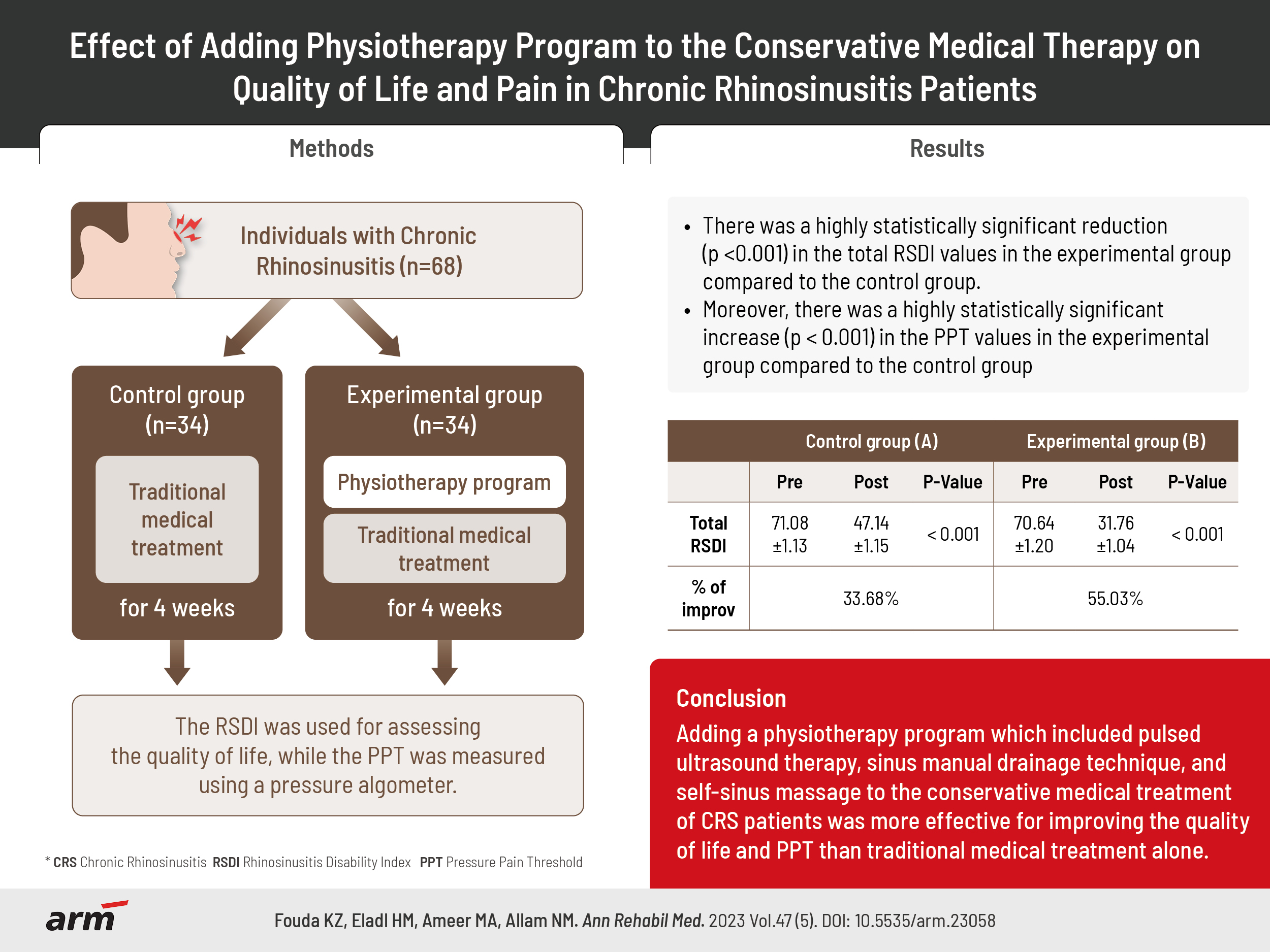
This study enrolled sixty-eight participants who suffering from CRS that have been clinically identified by an ear, nose, and throat (ENT) professional with the following criteria.
Inclusion criteria
Participants of both sexes, aged from 30 to 50 years, with a history of CRS lasting more than three months and clinical diagnostic criteria confirmation two or more main symptoms, or just one chief symptom (nasal blockage, pressure or soreness in the face, postnasal drip, and hyposmia) and two slight symptoms (headache, bad breath, exhaustion, tooth discomfort, and ear pain) and also confirmed by computed tomography (CT) scan outcomes [9].
Exclusion criteria
The exclusion criteria included the presence of any tumors or cysts (as proved by CT scan examination), nasal polyps, lesions on the face, illnesses, or allergies to the face, pregnancy, facial metal implants, previous surgery on the nose, and reduced heat perception (like uncontrolled type 2 diabetes), and deterioration of cognitive level.
Sample size calculation
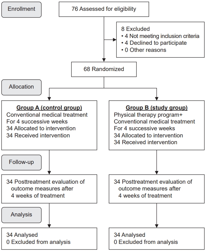
The sample size was calculated prior to the experiment to eliminate type II error. The calculations were performed using the statistical tool G*Power 3.1.9.4 at α=0.05, β=0.2, and effect size=0.75. It was determined that the necessary sample size was n=62. The sample size was increased to 68 participants to account for the drop-off as shown in Fig. 1.
Randomization
Sixty-eight CRS patients were sorted into one of two groups at random: control (group A) or experimental (group B). The randomization was carried out by a statistician who was not involved in the data gathering and who used a computer-generated random number list. Sealable, sequentially numbered opaque envelopes were utilized to assure the secret allocation. The first author opened the envelopes and began the treatment, as directed by the group assignment. The second author, who was not aware of the group assignment, got the outcome measures. Participants were blinded because those in the control group were referred by an ENT specialist and only had two encounters with the researcher, once for pretest evaluation and once for posttest evaluation four weeks later. The participants in the control group did not match those in the experimental groups. Furthermore, the experimental group’s physiotherapy sessions were private, separate, and held on different days and times. As a result, the participants have never met or know each other. In addition, the investigator ensures that participants in the study group are unaware of particular elements of the study.
Intervention
The patients were divided into two equal groups at random (n=34). Group A (the control group) received just prescription medicine from an ENT doctor. Group B (experimental group) was treated with medicine by an ENT specialist and involved in a program of physiotherapy that included: (1) US therapy: Using Sonoplus 490 from Enraf-Nonius, the participants were instructed to lie on their backs with the therapist standing at head height. The patients underwent pulsed US (duty cycle 50%) therapy for the maxillary and frontal sinuses at intensities of 1 and 0.5 W/cm2, respectively, and a 1 MHz collimating beam frequency with a 6:1 beam non-uniformity ratio, to deliver the US to the treatment area, A diminutive US applicator (0.8 cm2) with a 0.6 cm2 effective radiating area was utilized. The skin around the cheeks was utilized during application for the maxillary sinus and the forehead for the frontal sinus. Between the applicator and the skin, use an ultrasonic transmission gel, each maxillary sinus had a full contact approach for 5 minutes, while each frontal sinus received a 4 minutes full contact approach [10]. (2) Manual drainage techniques: It is performed from a supine lying position, while the therapist is seated behind the patient’s head. (3) Self-sinus massage technique: Participants were also urged to self-massage their frontal and maxillary sinuses at home while reclining twice daily, in the morning and evening. Table 1 has a full explanation of all manual approaches [15-17,20,21]. Before the trial began, a demonstration session was conducted to present participants in the experimental group with a detailed explanation of the manual therapy technique. The physiotherapy program was applied three times a week (day after day) for twelve therapy sessions. The session started with US at the frontal sinuses then the maxillary sinuses followed immediately by manual drainage technique, the session lasted approximately 30 minutes.
Outcome measures
The measurable outcomes were assessed at baseline to 4 weeks after treatment, directly after the completion of the twelve sessions of the treatment program by the author, who was blinded to the allocation. The participants were asked not to use any topical or systemic nasal drugs in the previous 24 hours before the baseline examination. The rhinosinusitis disability index (RSDI) was the primary outcome measure, while the PPT was the secondary outcome measure.
RSDI
The Arabic version of RSDI was used to evaluate the influence of CRS on patients’ QoL. It is a precise, validated, and reliable (Cronbach’s alpha=0.97) questionnaire for use with Arabic-speaking patients suffering from rhinosinusitis [22]. It includes 30 elements related to sinus and nasal symptoms that can result in distinct limitations on daily activities. The RSDI is divided into three areas: emotional (10 items), physical (11 items), and functional (9 items). Each item is assessed on a five-point Likert scale, between never (scored as 0) to always (scored as 4). The possible overall score runs from 0 to 120, with higher values indicating lower health-related QoL and higher levels of impairment [23,24].
PPT
The PPT in the target sinus, which is the least pressure that causes pain in tissue trigger sites [25], was measured using the FPX 25 Digital Algometer (Wagner Instruments). The measuring unit was calibrated as kg/cm2 (capacity/graduation=10×0.01 kgf). Pressure algometry is a valid and reliable method of measuring pain in the muscles, fascia, joints, tendons, ligaments, and periosteum [26,27]. The patient was positioned supine, and the 1 cm2 rubber-tipped end of the algometer which placed vertically to the skin surface over the predetermined areas in the frontal sinus (between the bridge of the nose and the inner side of the upper eyelid) and maxillary sinus (just below the cheekbones), respectively. A constant, mild pressure was administered until the patient felt pain for the first time and answered with “now.” After removing the algometer, the value was recorded as the PPT for that sinus.
Statistical analysis
Descriptive statistics and an independent t-test were applied to compare the characteristics of the patients in both groups. The Wilcoxon signed rank test and Mann–Whitney u-test were used to compare the RSDI scores within and between groups. While the dependent t-test and independent t-test were used to compare the PPT scores within and between groups. The level of significance was fixed at alpha<0.05.
RESULTS
The patients comprised 31 male (45.59%) and 37 female (54.41%) with a mean age of 38.40 years and a body mass index of 26.79 kg/m2. The independent t-test revealed that there were no significant differences (p>0.05) among the groups regarding patients’ characteristics as shown in Table 2.
Wilcoxon signed-rank test indicated that there was a significant reduction (p<0.001) in RSDI values for both groups when comparing the pretreatment values vs. the posttreatment values for each group as shown in Table 3. For control group (A), the physical disability index value decreased from 32.47±1.50 before treatment to 20.88±1.27 after treatment with a mean difference of 11.58 and the percentage of improvement was 35.66%, the functional disability index value also decreased from 19.03±1.44 before treatment to 12.88±1.34 after treatment with a mean difference of 6.14 and the percentage of improvement was 32.28%, the emotional disability index value decreased from 19.58±1.57 before treatment to 13.38±0.81 after treatment with a mean difference of 6.20 and the percentage of improvement was 31.66%, the total RSDI value decreased from 71.08±1.13 before treatment to 47.14±1.15 after treatment with a mean difference of 23.94 and the percentage of improvement was 33.68%.
While for the experimental group (B), the physical disability index value reduced from 32.88±1.37 before treatment to 14.17±1.33 after treatment with a mean difference of 17.91 and the percentage of improvement was 55.82%, the functional disability index value also reduced from 18.70±1.36 before treatment to 8.76±0.74 after treatment with a mean difference of 9.94 and the percentage of improvement was 53.15%, the emotional disability index value reduced from 19.82±1.48 before treatment to 8.82±0.86 after treatment with a mean difference of 11.00 and the percentage of improvement was 55.49%, the total RSDI value reduced from 70.64±1.20 before treatment to 31.76±1.04 after treatment with a mean difference of 38.88 and the percentage of improvement was 55.03%. The results revealed that the experimental group had a much higher percentage of improvement in RSDI values than the control group as shown in Table 3.
The Mann–Whitney U-test showed no evidence of a significant difference (p>0.05) in RSDI values between both groups at the pretest conditions, while for the posttest conditions, there was a highly statistically significant decrease (p<0.001) in RSDI values in the experimental group in comparison to the control group as shown in Table 3. Moreover, there was a statistically significant difference (p<0.001) between both groups when comparing the change of the pre-post data values.
For control group (A), the change between the pretest and posttest values for physical disability index was 11.58±1.33, for functional disability index was 6.14±0.43, for emotional disability index was 6.20±1.12, for the total RSDI was 23.94±0.95. While for the experimental group (B), the change between the pretest and posttest values for the physical disability index was 17.91±1.11, for the functional disability index was 9.94±0.91, for the emotional disability index was 11.00±0.77, for the total RSDI was 38.88±0.67 as shown in Table 3.
The dependent t-test showed that there was a significant increase (p<0.001) in PPT values for both groups when comparing the pretreatment values vs. the posttreatment values for each group. For control group (A), the PPT of the right frontal sinus increased from 1.45±0.16 before treatment to 2.06±0.16 after treatment with a mean difference of 0.61 and the percentage of improvement was 42.06%, while the left frontal sinus PPT increased from 1.43±0.14 before treatment to 2.03±0.13 after treatment with a mean difference of 0.60 and the percentage of improvement was 41.95%. The PPT of the right maxillary sinus also increased from 1.90±0.11 before treatment to 2.57±0.10 after treatment with a mean difference of 0.67 and the percentage of improvement was 35.26%, while the left maxillary sinus PPT increased from 1.86±0.06 before treatment to 2.52±0.09 after treatment with a mean difference of 0.66 and the percentage of improvement was 35.48%.
While for the experimental group (B), the PPT of the right frontal sinus increased from 1.48±0.11 before treatment to 3.07±0.17 after treatment with a mean difference of 1.59 and the percentage of improvement was 107.43%, while the left frontal sinus PPT increased from 1.44±0.08 before treatment to 3.05±0.13 after treatment with a mean difference of 1.60 and the percentage of improvement was 111.80%. The PPT of the right maxillary sinus also increased from 1.87±0.10 before treatment to 3.21±0.15 after treatment with a mean difference of 1.33 and the percentage of improvement was 71.65%, while the left maxillary sinus PPT increased from 1.84±0.07 before treatment to 3.17±0.11 after treatment with a mean difference of 1.32 and the percentage of improvement was 72.47%. According to the findings, the experimental group’s PPT values improved by a much greater percentage than those of the control group as shown in Table 4.
Independent t-test showed no significant difference (p>0.05) in PPT values between both groups at the pretest conditions, while for the posttest conditions, there was a highly statistically significant increase (p<0.001) in PPT values in the experimental group compared to the control group. Furthermore, there was a statistically significant difference (p<0.001) between both groups when comparing the change of the pre-post data values.
For control group (A), the change between the pretest and posttest values of the right frontal sinus PPT was 0.61±0.16 and it was 0.60±0.14 for the left frontal sinus PPT. Whereas, the right maxillary sinus PPT was 0.67±0.06 and it was 0.66±0.09 for the left maxillary sinus PPT. While for the experimental group (B) the change between the pretest and posttest values of the right frontal sinus PPT was 1.59±0.16 and it was 1.60±0.14 for the left frontal sinus PPT. Whereas, the right maxillary sinus PPT was 1.33±0.05 and it was 1.32±0.08 for the left maxillary sinus PPT as shown in Table 4.
DISCUSSION
The present study’s findings showed that the experimental group which received the physical therapy program in conjunction with conventional medical treatment demonstrated a highly statistically significant (p<0.001) enhancement in the measured outcomes after 4 weeks of treatment when compared to the control group that received only conservative medical therapy.
The current study’s findings supported the authors’ hypothesis that adding physical therapy programs including US therapy and manual therapy program to the traditional medical treatment was more effective than conventional medical treatment alone in treating patients with CRS in terms of QoL and PPT. QoL and pain enhancement can be attributed to a proposed strategy for the US that involves the breakdown of the biofilm structure of the bacterial population. The US was reported to reduce bacterial load by destroying biofilms [12]. Moreover, purulent discharge was frequently seen during or right after receiving US treatment. This could be because the US delivered mechanical energy to separate the purulent material from the sinus walls, relieving pressure and pain [28].
Pulsed US therapy for CRS patients has been shown to be significantly more efficient when combined with antibiotics, which can significantly reduce bacterial viability [28-31]. Additionally, 57 patients with CRS were successfully treated with low-intensity pulsed US. The investigators reported that the majority of both major and minor symptoms showed significant improvements following pulsed US therapy [32].
Other studies by Ansari et al. [8] showed a significantly larger decline in CRS symptoms following treatment with the US. Furthermore, when comparing pulsed and continuous therapeutic US, they did not identify any significant differences in outcomes between the two groups [9], however, pulsed US mode minimizes thermal activities by giving the coupling medium time to dissipate heat during treatment [33]. Another study included 20 patients who received six sessions of US three days per week. The severity of global sinonasal symptoms was evaluated after treatment using a 6-cm visual analog and the Sino-Nasal Outcome Test. Following treatment with the US, the patient’s total severity assessment scores improved [34].
One more case series study incorporates manual therapy into the overall management of CRS symptoms, and they found that patients who received a combination of local and regional manual therapy procedures, improved in all measured outcomes. There was a significant reduction in craniofacial pain and an increase in PPTs over four precise sinus points, as well as a reduction in the severity of symptoms. These findings seemed to compare more positively with outcomes seen in similar patients treated with antibiotics or endoscopic surgery [15].
The PPT was found to be significantly increased, as measured by pressure algometry on the frontal and maxillary sinuses. The positive effect could be attributed to the thermal effects produced at the sinuses as a result of the manual therapy intervention, which assisted in draining the excess secretions that cause inflammation to the adjacent lymph nodes. This technique, in turn, helped to reduce sinus inflammation and pain [35]. In agreement with our findings, Ahmadi et al. [20] indicated that massage therapy can be incorporated into an exercise program as a treatment modality in patients with CRS, based on the findings of their study, which revealed that a special face massage therapy protocol can relieve facial congestion and tenderness in CRS patients.
A five-session study combining US and shortwave diathermy interventions using manual drainage procedures and suboccipital release showed significant improvements in patients with chronic sinusitis. However, when compared to the shortwave diathermy group, the US therapy group experienced earlier and more rapid symptom reduction. As a result, the study recommended that US therapy would be applied as a treatment protocol for patients suffering from chronic sinusitis [19]. Thus, a novel approach to treating chronic sinusitis is currently being developed, which improves medical management and reduces antibiotic resistance, hence reducing the need for surgical surgery.
CRS has a similar negative influence on health as angina, chronic obstructive lung disorders, congestive heart failure, and low back pain [36], Surgery is typically the next step after oral, topical, and antibiotic therapy. The establishment of a new paradigm in the treatment of CRS could result from the effective deployment of a physical therapy program. To the best of the authors’ knowledge, this is the initial research that evaluates the effect of adding US therapy to manual techniques using frontal, nasal, and maxillary sinus drainage, in addition to self-massage techniques and routine medical treatment.
This study was limited by the evaluation of improvement by CT in the area of para-nasal sinus, which might be a beneficial tool for the assessment of the anatomy and extent of improvement, thus more studies are recommended to evaluate the improvement by more additional assessment tools (sinuscopy and CT). In addition, only the short-term effect of the addition of a manual physical therapy program was evaluated, thus, long-term follow-up should be considered in further studies. Moreover, certain parameters of US were used in this study, different parameters will be recommended in future studies to assess their effects. In addition, further research is required to determine if manual techniques or US therapy plays a more important role in treating chronic instances of rhinosinusitis because the mixed physiotherapy program may not make clear which percentage of improvement was due to US therapy or manual techniques, so further study is needed to clarify this point. Furthermore, the sham US, sham sinus manual drainage techniques, and sham self-sinus massage weren’t be given to the control group therefore future research should incorporate this type of intervention to improve study blinding. Also, this study was limited to a certain age group, therefore, more studies are recommended to assess the effect of different treatment periods of application on different categories of age.
In conclusion, according to the findings of this study, incorporating a physical therapy program that included pulsed US therapy, sinus manual drainage techniques, and self-sinus massage into conservative medical treatment was more effective than conservative medical treatment alone in improving QoL and PPT in CRS patients.