• KARM
  • Contact us
  • E-Submission
ABOUT
ARTICLE TYPES
BROWSE ARTICLES
AUTHOR INFORMATION

Articles

Original Article

Changes in Activation of Serratus Anterior, Trapezius and Latissimus Dorsi With Slouched Posture

Annals of Rehabilitation Medicine 2016;40(2):318-325.
Published online: April 25, 2016

Department of Rehabilitation Medicine, CHA Bundang Medical Center, CHA University, Seongnam, Korea.

Corresponding author: Kyunghoon Min. Deprtment of Rehabilitation Medicine, CHA Bungdang Medical Center, CHA University, 59 Yatap-ro, Bundang-gu, Seongnam 13496, Korea. Tel: +82-31-780-6287, Fax: +82-31-780-6206, minkh@chamc.co.kr
• Received: July 21, 2015   • Accepted: September 8, 2015

Copyright © 2016 by Korean Academy of Rehabilitation Medicine

This is an open-access article distributed under the terms of the Creative Commons Attribution Non-Commercial License (http://creativecommons.org/licenses/by-nc/4.0) which permits unrestricted noncommercial use, distribution, and reproduction in any medium, provided the original work is properly cited.

  • 11,654 Views
  • 176 Download
  • 29 Web of Science
  • 31 Crossref
  • 31 Scopus
prev next
  • Objective
    To compare quantitative muscle activation between erect and slouched sitting postures in the muscles around the scapula, and to investigate the correlation between the angle of thoracic kyphosis and the alteration of muscle activity depending on two different sitting postures.
  • Methods
    Ten healthy males participated in the study. Unilateral surface electromyography (SEMG) was performed for serratus anterior, middle trapezius (MT), and lower trapezius (LT), which are scapular stabilizer muscles, as well as latissimus dorsi. Participants elevated their shoulders for 3 seconds up to 90° abduction in the scapular plane, tilting 30° anterior in the coronal plane. They were told to hold the position for 10 seconds and voluntary isometric contractions were recorded by SEMG. These movement procedures were conducted for three times each for erect and slouched sitting postures and data were averaged.
  • Results
    Activities of MT and LT increased significantly more in the slouched sitting posture than in the erect one. There was no significant correlation between kyphotic angle and the area under curve of each muscle.
  • Conclusion
    Because MT and LT are known as prime movers of scapular rotation, the findings of this study support the notion that slouched sitting posture affects scapular movement. Such scapular dyskinesis during arm elevation leads to scapular stabilizers becoming overactive, and is relevant to muscle fatigue. Thus, slouched sitting posture could be one of the risk factors involved in musculoskeletal pain around scapulae.
Slouched posture is defined as a relaxed sitting posture with a flexed thoracic and lumbar spine. Such a posture is commonly encountered in our daily activities [1]. Those who use a computer often and who sit for a long time have been reported to assume this slouched position with greater frequency [2]. The posture is related to musculoskeletal pain [3], and leads to cervical and thoracic muscles becoming overactive along with sprain of the iliolumbar ligament [45].
In addition, relaxed sitting has resulted in the kinematics of scapulae to become changed during arm elevation, with the reduction of posterior tipping and lateral rotation of scapulae [6]. Thoracic kyphosis is caused by a slouched posture and spinal inclination, and is one of the risk factors for a limited shoulder range of motion [7]. When subjects bend forward, scapulae become more protracted and rotated downwardly, causing rotator cuff muscles to become compressed in the subacromial space. Shoulder impingement syndrome is known to be associated with inappropriate scapular positioning [8]. Briefly, shoulder elevation occurs naturally with the thoracic spine in an extended position and slouched posture limits this motion.
Scapular position and movement to the thorax are critical for normal glenohumeral function and facilitates optimal shoulder movements [91011]. The scapula provides a base for glenohumeral mobility and surrounding muscles play a role in scapula stabilization. During overhead arm elevation movements at the glenohumeral joint, it is greatly important that the scapular-stabilizing musculature be strong enough to properly sustain the scapula. The muscles attached on the medial aspect of the scapula are the key muscles for stabilization. These include the middle and lower trapezius, rhomboid major and minor, and serratus anterior [12]. Previous studies have investigated the effect of scapular stabilizer muscles according to sitting posture [61314], but no study to date reports quantitatively on muscle activities with electromyography (EMG).
We hypothesized that a slouched sitting posture may induce inappropriate activation patterns of key muscles for scapular stabilization such as the serratus anterior (SA), mid-trapezius (MT), and low-trapezius (LT). In consideration of its origin and insertion between the thoracolumbar spine and humerus, latissimus dorsi (LD) was also expected to be affected, and was therefore included in this study. This study aims to compare quantitative activation of those four muscles between upright and slouched sitting postures, and to investigate the correlation between the angle of thoracic kyphosis and the alteration of muscle activities.
Subjects
A prospective cross-sectional study was conducted from February 2015 to May 2015. Ten healthy males participated. Participants were enrolled if they were in good health and had no shoulder or neck problems. Those with a limited range of shoulder motion; surgical interventions within 6 months in the dominant shoulder; complaint of pain in the neck and shoulder; and having a temporary external pacemaker, were excluded. The study protocol was approved by the Institutional Review Board of the CHA Bundang Medical Center, and all participants provided written informed consent.
Methods

Evaluation of the shoulder pain and function

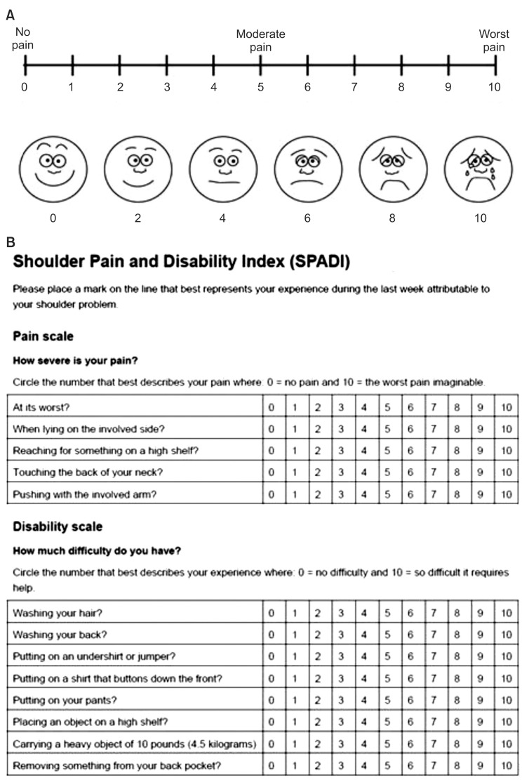
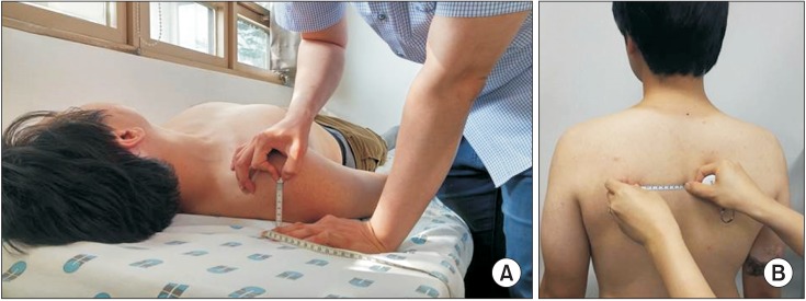
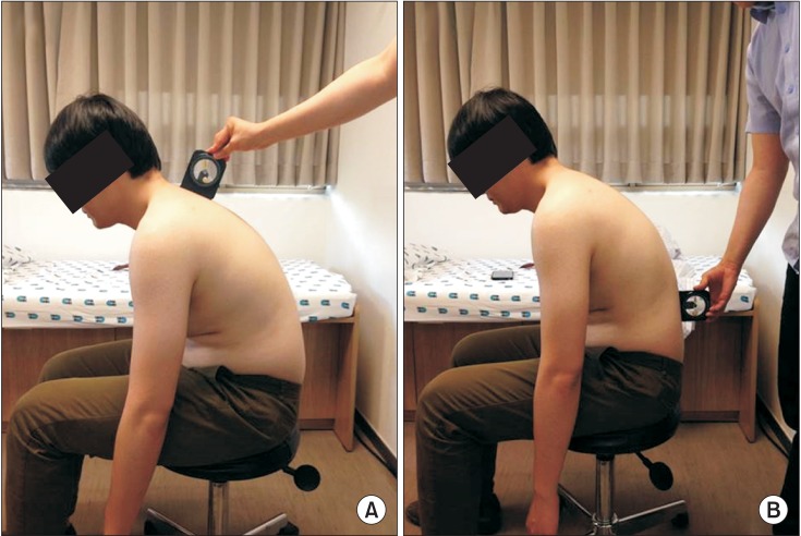
Subjects were evaluated for shoulder pain intensity and function using the Numerical Rating Scale (NRS) and the Shoulder Pain and Disability Index (SPADI) (Fig. 1). Two clinical tests were then performed for the assessment of scapular positioning [8] (Fig. 2). First, subjects were instructed to relax in the supine position and the distance between the posterior border of the acromion and the table was measured. The assessor also measured the distance between acromial posterior border and the table on bilateral sides. Second, the medial scapular border and the fourth thoracic spinous process were palpated and the distance between them was measured while the subject was in a standing position. The above two clinical measurements were done in two postures: the relaxed status and the shoulder retracted position, for which the participant was instructed to move both shoulders as far backward as possible.

Procedure

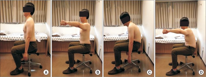
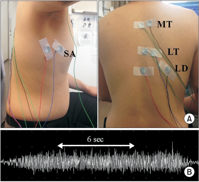
Subjects were seated in the trunk upright posture. They contracted their shoulder muscles with 90° abduction in the scapular plane while tilting 30° anteriorly in the coronal plane for 3 seconds, and were told to hold the position for 10 seconds [6] (Fig. 3). They repeated this procedure three times with 10 seconds of rest. Before collecting the data, three trials of the movements were performed for the participants to be familiar with the movements. The activities of muscles were measured quantitatively by calculating the area under curve (mV·ms) of the middle 6 seconds during isometric contraction (Fig. 4). Next, the subjects were instructed to maintain a slouched posture as long as they could. Angles between T1/T2 and T12/L1 were calculated for kyphotic angle with an inclinometer [15] (Fig. 5). They repeated the previous procedure in a slouched posture after 3 minutes rest. All measurements were performed by a single experienced physiatrist.

SEMG recording

Unilateral surfaces EMG (SEMG) was performed in a similar manner to previous studies [16]. Four muscles were recorded: SA, MT, LT, and LD (Fig. 4). Active electrodes were placed at the muscle belly of 6th rib for SA [17], 20% medial to the midpoint between the medial border of the scapular and the T4 for MT [18], 33% medial to the midpoint between the medial scapular and the T8 for LT [18], and 4 cm below the inferior tip of the scapula for LD [1920]. The reference electrodes were attached to the spinous process on the same level as the active electrodes. The reference electrode for the SA was placed at the mid-axillary line of the 6th rib. The ground electrode was positioned at the clavicle [21]. The sites of electrode attachment were prepared using fine sandpaper and 70% isopropyl alcohol. In some cases, when required, excess hair was shaved. Small disposable electrodes (Ag/AgCl discs, 10-mm diameter; CareFusion, San Diego, CA, USA) in a bipolar configuration were used to record the SEMG of each muscle. The electrodes were attached with paper tape during the procedure to avoid any movement of the electrodes.

Data analysis

The SEMG signals were collected with an eight-channel EMG Nicolet EDX (Natus Medical Inc., San Carlos, CA, USA), band-pass filtered from 20 to 250 Hz using fullwave rectification [16]. As the area under curve (AUC) was measured, no smoothing was done. EMG signals were digitized, sampled at frequency of 1,200 Hz, and stored using Viking electrodiagnostic software (ver. 20.0.34; Natus Medical Inc.) for data processing and analysis. During data collection, raw EMG recordings were monitored. The AUC (mV·ms) of the middle 6 seconds during isometric contraction was calculated for the quantitative measurement.

Statistical analysis

Data were normalized by measuring the AUC at 3 seconds after onset of steady isometric voluntary contraction for 6 seconds. The acquired AUC was calculated into percentiles for each patient and then statistically analyzed. To compensate for the differences in individual muscle mass and activation patterns, datasets were normalized by dividing each recording of slouched posture with mean recorded values of the upright posture for each corresponding muscle. The Wilcoxon signed-rank test was used to assess the differences of the AUC of the EMG signal according to the sitting postures and the asymmetry between right and left shoulders in acromion and scapular movement tests. The relationship between the kyphotic angle and muscle activation was analyzed using the Spearman correlation test. The results are reported as the mean±standard deviation in the text, and as mean and standard error of the mean in the figures. All statistical analyses were performed with the statistical software package SPSS ver. 21.0 (SPSS Inc., Chicago, IL, USA). The level of statistical significance employed was p<0.05.
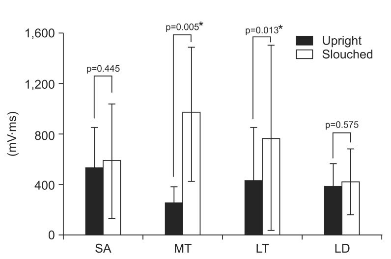
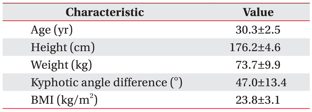
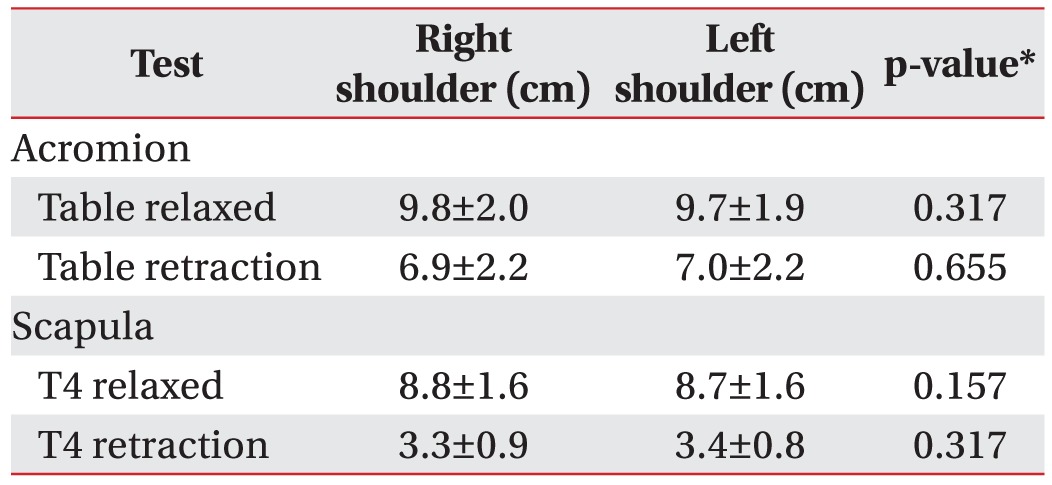
Ten healthy volunteers completed all the procedures. They were all males and their demographic data are shown in Table 1. All subjects that had no shoulder pain were assessed with NRS and SPADI. There was no asymmetry between right and left acromion and scapular movements in the two clinical tests (Table 2). Significant differences in MT and LT were found between two positions according to SEMG data. MT and LT activity were significantly increased in the slouched sitting position (972.9±521.1 and 770.9±737.1, respectively) as compared to the erect sitting group (262.4±122.4 and 435.8±420.4, respectively) (Table 3, Fig. 6). For SA and LD, there was no difference in the muscle activities between the two positions.
There was no significant correlation between the kyphotic angle and AUC of each muscle (Table 4).
This study aimed to investigate the effects of the slouched sitting posture on the muscles surrounding the scapula during arm elevation. The results indicate that the slouched sitting posture leads to more activation of MT and LT (Table 3). These two muscles are part of the group of scapular stabilizers along with rhomboid major, rhomboid minor and serratus anterior. MT is a scapular retractor and LT upward rotates and depresses the scapula [12]. Chronic hyperactivation and scapular dyskinesis of MT and LT may contribute to shoulder pain. A previous study reported that shoulder impingement syndrome patients showed increased upper and lower trapezius muscle activity during humeral elevation [22]. Another study indicated that hyperactivation of the muscle to maintain a certain posture can result in fatigue of the associated muscle and may induce repetitive injury in the shoulder pain syndrome [23]. The effects of muscle fatigue and external load on scapular kinematics during humeral elevation have also been reported in scapular dyskinesis, which is associated with shoulder pain [24]. In short, as shown in our study, a continuous slouched posture can lead to chronic hyperactivation of LT and MT, resulting in muscle fatigue. Our study supports the previously held notion that slouched posture can be one of the pain sources in the shoulder.
No difference in the activation of SA and LD was noticed in this study between the erect and slouched sitting positions. LD inserts along the intertubercular groove of the humerus and functionally works as a shoulder joint muscle, assisting in adduction, extension and medial rotation [25]. Considering the bridging of LD between the thoracolumbar spine and the humerus bypassing the scapula, the direct influence of different sitting positions during arm elevation on LD might not be significant. In addition, this study only assessed humeral abduction, which might have little effect on LD. As for SA, according to a previous study, there was no change in the activity of SA depending on the flexed head position, which is consistent with our results [26]. A different study reported that the activity of SA decreased during humeral elevation with a forward head posture [27]. This difference may be due to the different study methods and small sample sizes of both studies, and they remain to be validated in studies with larger samples sizes.
One limitation of the surface electrode recording technique is the cross-talk phenomenon that obtains signals from proximal irrelevant muscles. In our study, the surface electrodes of MT and LT may have recorded signals from the rhomboid muscle. However, the majority of the signals obtained in the surface electrodes are expected to be from the most superficial muscles, MT and LT, and not the rhomboids. Further studies acquiring signals from the needle EMG during isometric contractions may further clarify the result.
As a pilot study, this study is also limited by the small sample size. Another limitation was that the muscle activity was assessed in only the arm elevation in the scapular plane. The shoulder joint shows 3-dimensional movement and arm elevation does not represent the whole picture. Furthermore, only static postures were investigated in this study. Real life postures are dynamic and may have different activation patterns. Finally, the actual clinical correlation of slouched posture to significant shoulder pain has not been well studied. Future studies involving more participants, access of all shoulder movements, integration of dynamic components of sitting posture, and prospective cohorts of volunteers with slouched posture, may further strengthen the result and application of this study.
According to our investigation, slouched sitting posture leads MT and LT to become hyperactive. Because MT and LT are known as prime movers of scapular rotation, the findings support the notion that abnormal posture of the spine, especially a slouched sitting position, is a potential source of pain originating from scapular dyskinesis. Therefore, spine structure needs to be evaluated in patients with shoulder and back pain, and therapeutic intervention relating to slouched posture would be helpful for pain control.

CONFLICT OF INTEREST: No potential conflict of interest relevant to this article was reported.

  • 1. Dolan KJ, Green A. Lumbar spine reposition sense: the effect of a 'slouched' posture. Man Ther 2006;11:202-207.
  • 2. Kleine BU, Schumann NP, Bradl I, Grieshaber R, Scholle HC. Surface EMG of shoulder and back muscles and posture analysis in secretaries typing at visual display units. Int Arch Occup Environ Health 1999;72:387-394.
  • 3. Mirbagheri SS, Rahmani-Rasa A, Farmani F, Amini P, Nikoo MR. Evaluating kyphosis and lordosis in students by using a flexible ruler and their relationship with severity and frequency of thoracic and lumbar pain. Asian Spine J 2015;9:416-422.
  • 4. Snijders CJ, Hermans PF, Niesing R, Jan Kleinrensink G, Pool-Goudzwaard A. Effects of slouching and muscle contraction on the strain of the iliolumbar ligament. Man Ther 2008;13:325-333.
  • 5. Caneiro JP, O'Sullivan P, Burnett A, Barach A, O'Neil D, Tveit O, et al. The influence of different sitting postures on head/neck posture and muscle activity. Man Ther 2010;15:54-60.
  • 6. Finley MA, Lee RY. Effect of sitting posture on 3-dimensional scapular kinematics measured by skinmounted electromagnetic tracking sensors. Arch Phys Med Rehabil 2003;84:563-568.
  • 7. Imagama S, Hasegawa Y, Wakao N, Hirano K, Muramoto A, Ishiguro N. Impact of spinal alignment and back muscle strength on shoulder range of motion in middle-aged and elderly people in a prospective cohort study. Eur Spine J 2014;23:1414-1419.
  • 8. Nijs J, Roussel N, Vermeulen K, Souvereyns G. Scapular positioning in patients with shoulder pain: a study examining the reliability and clinical importance of 3 clinical tests. Arch Phys Med Rehabil 2005;86:1349-1355.
  • 9. Brumitt J. Scapular-stabilization exercises: early-intervention prescription. Athl Ther Today 2006;11:15-18.
  • 10. Voight ML, Thomson BC. The role of the scapula in the rehabilitation of shoulder injuries. J Athl Train 2000;35:364-372.
  • 11. Ludewig PM, Reynolds JF. The association of scapular kinematics and glenohumeral joint pathologies. J Orthop Sports Phys Ther 2009;39:90-104.
  • 12. Escamilla RF, Yamashiro K, Paulos L, Andrews JR. Shoulder muscle activity and function in common shoulder rehabilitation exercises. Sports Med 2009;39:663-685.
  • 13. Bullock MP, Foster NE, Wright CC. Shoulder impingement: the effect of sitting posture on shoulder pain and range of motion. Man Ther 2005;10:28-37.
  • 14. Kanlayanaphotporn R. Changes in sitting posture affect shoulder range of motion. J Bodyw Mov Ther 2014;18:239-243.
  • 15. Lewis JS, Valentine RE. Clinical measurement of the thoracic kyphosis. A study of the intra-rater reliability in subjects with and without shoulder pain. BMC Musculoskelet Disord 2010;11:39.
  • 16. Lee HS, Shim JS, Lee ST, Kim M, Ryu JS. Facilitating effects of fast and slope walking on paraspinal muscles. Ann Rehabil Med 2014;38:514-522.
  • 17. Cools AM, Witvrouw EE, De Clercq GA, Danneels LA, Willems TM, Cambier DC, et al. Scapular muscle recruitment pattern: electromyographic response of the trapezius muscle to sudden shoulder movement before and after a fatiguing exercise. J Orthop Sports Phys Ther 2002;32:221-229.
  • 18. Holtermann A, Roeleveld K, Mork PJ, Gronlund C, Karlsson JS, Andersen LL, et al. Selective activation of neuromuscular compartments within the human trapezius muscle. J Electromyogr Kinesiol 2009;19:896-902.
  • 19. Park SY, Yoo WG. Differential activation of parts of the latissimus dorsi with various isometric shoulder exercises. J Electromyogr Kinesiol 2014;24:253-257.
  • 20. Beaudette SM, Unni R, Brown SH. Electromyographic assessment of isometric and dynamic activation characteristics of the latissimus dorsi muscle. J Electromyogr Kinesiol 2014;24:430-436.
  • 21. Noguchi M, Chopp JN, Borgs SP, Dickerson CR. Scapular orientation following repetitive prone rowing: implications for potential subacromial impingement mechanisms. J Electromyogr Kinesiol 2013;23:1356-1361.
  • 22. Ludewig PM, Cook TM. Alterations in shoulder kinematics and associated muscle activity in people with symptoms of shoulder impingement. Phys Ther 2000;80:276-291.
  • 23. McLean L. The effect of postural correction on muscle activation amplitudes recorded from the cervicobrachial region. J Electromyogr Kinesiol 2005;15:527-535.
  • 24. Poppen NK, Walker PS. Normal and abnormal motion of the shoulder. J Bone Joint Surg Am 1976;58:195-201.
  • 25. Bagg SD, Forrest WJ. A biomechanical analysis of scapular rotation during arm abduction in the scapular plane. Am J Phys Med Rehabil 1988;67:238-245.
  • 26. Ludewig PM, Cook TM. The effect of head position on scapular orientation and muscle activity during shoulder elevation. J Occup Rehabil 1996;6:147-158.
  • 27. Weon JH, Oh JS, Cynn HS, Kim YW, Kwon OY, Yi CH. Influence of forward head posture on scapular upward rotators during isometric shoulder flexion. J Bodyw Mov Ther 2010;14:367-374.
Fig. 1

The measurement of Shoulder Pain and Disability Index. (A) Numeric rating scale, (B) Shoulder Pain and Disability Index.

arm-40-318-g001.jpg
Fig. 2

Two clinical tests for the assessment of scapular positioning: (A) the distance between the posterior border of the acromion and the table in the supine position, (B) the distance between the medial border and the fourth thoracic spinous process.

arm-40-318-g002.jpg
Fig. 3

Upright sitting and slouched sitting position: (A) resting upright sitting position, (B) upright sitting position with shoulder abduction, (C) resting slouched sitting position, and (D) slouched sitting position with shoulder abduction.

arm-40-318-g003.jpg
Fig. 4

Recording sites of surface electromyography (EMG) and measurement timing of muscle activation. (A) Serratus anterior (SA), middle trapezius (MT), lower trapezius (LT) and latissimus dorsi (LD). (B) Area under curve (mV·ms) was measured for 6 seconds.

arm-40-318-g004.jpg
Fig. 5

Measurement sites of thoracic kyphosis. (A) The level of 1st and 2nd thoracic spine. (B) The level of 12th thoracic spine and 1st lumbar spine. Thoracic kyphosis=(A) angle+(B) angle.

arm-40-318-g005.jpg
Fig. 6

Differences of muscle activities between upright and slouched sitting positions (n=10). Values are presented as mean±standard deviation. SA, serratus anterior; MT, middle trapezius; LT, lower trapezius; LD, latissimus dorsi. *p<0.05 by Wilcoxon signed-rank test.

arm-40-318-g006.jpg
Table 1

Baseline characteristics of subjects (n=10)

Values are presented as mean±standard deviation.

BMI, body mass index.

arm-40-318-i001.jpg
Table 2

Descriptive statistics of the outcome on the two clinical tests for the assessment of scapular positioning (n=10)

Values are presented as mean±standard deviation.

*p<0.05 by Wilcoxon signed-rank test.

arm-40-318-i002.jpg
Table 3

Differences of muscle activities between the upright and slouched sitting position (n=10)

Values are presented as mean±standard deviation.

The slouched (%) indicates mean±standard deviation for the normalized data calculated for the slouched posture. SA, serratus anterior; MT, middle trapezius; LT, lower trapezius; LD, latissimus dorsi.

*p<0.05 by Wilcoxon signed-rank test.

arm-40-318-i003.jpg
Table 4

Correlation between kyphotic angle and muscle activation difference (n=10)

The (%) indicates mean±standard deviation for the normalized data calculated for the slouched posture.

SA, serratus anterior; MT, middle trapezius; LT, lower trapezius; LD, latissimus dorsi.

*p<0.05 by Spearman correlation analysis.

arm-40-318-i004.jpg

Figure & Data

References

    Citations

    Citations to this article as recorded by  
    • Influence of pelvic position on shoulder range of motion
      Bishoy S. Lobbos, Mohamed M. M. Essa, Alaaeldin Khaireldin, Mohamed Y. Gamal El-Din, Phillips Rizkallah, Heba Allah Samy
      BMC Musculoskeletal Disorders.2025;[Epub]     CrossRef
    • Comparative Study on Scapular Alignment and Neck and Shoulder Muscle Strength in Subjects with Forward Head Posture and Round Shoulder Posture
      Kyoung-Yeol Jeong, Tae-Gyu Kim, Il-Young Yu, Soo-Yong Kim
      The Korean Journal of Sports Medicine.2025; 43(1): 13.     CrossRef
    • The Correlation Between Cervical Proprioception and Scapular Dyskinesis in Patients With Neck Pain: A Case-Control Study
      Richa R Bisen, Pranaya D Kadam, Annamma Varghese, Rahul Bisen
      Cureus.2024;[Epub]     CrossRef
    • Targeted Motor Control Considering Sternal Position Improves Spinal Alignment in Pregnant Women at Risk for Preterm Birth with Low Back Pain
      Arkadiusz Żurawski, Sun Ha, Grzegorz Świercz, Olga Adamczyk Gruszka, Wojciech Kiebzak
      Journal of Clinical Medicine.2024; 13(24): 7661.     CrossRef
    • Effect of Scapular Stabilizer Muscles Strength on Scapular Position
      Mehmet Micoogullari, S. Fatma Uygur, H. Baran Yosmaoglu
      Sports Health: A Multidisciplinary Approach.2023; 15(3): 349.     CrossRef
    • Poor Sit-to-Stand Performance in Adolescent and Young Adult Patients with Sarcoma
      Savannah V. Wooten, Behrang Amini, J. Andrew Livingston, Michelle A.T. Hildebrandt, Joya Chandra, Susan C. Gilchrist, Michael Roth, Eugenie Kleinerman
      Journal of Adolescent and Young Adult Oncology.2023; 12(6): 821.     CrossRef
    • Analysis of abnormal muscle activities in patients with loss of cervical lordosis: a cross-sectional study
      Jiwoon Lim, Dajeong Lee, Sangyoung Kim, Seungeun Lee, Ju Seok Ryu
      BMC Musculoskeletal Disorders.2023;[Epub]     CrossRef
    • Effects of Alignment of the Thoracic Spine and Ankle Joint on Muscle Activation During Sling Exercise
      Hojin Shin, Gyeongseop Sim
      Physical Therapy Rehabilitation Science.2023; 12(3): 340.     CrossRef
    • Effect of Rounded and Hunched Shoulder Postures on Myotonometric Measurements of Upper Body Muscles in Sedentary Workers
      Rama Krishna Reddy Guduru, Aurelijus Domeika, Auksė Domeikienė
      Applied Sciences.2022; 12(7): 3333.     CrossRef
    • Intramuscular Neural Distribution of the Serratus Anterior Muscle: Regarding Botulinum Neurotoxin Injection for Treating Myofascial Pain Syndrome
      Kyu-Ho Yi, Ji-Hyun Lee, Hee-Jin Kim
      Toxins.2022; 14(4): 271.     CrossRef
    • Guidance to trigger point injection for treating myofascial pain syndrome: Intramuscular neural distribution of the quadratus lumborum
      Kyu‐Ho Yi, Kyu‐Lim Lee, Ji‐Hyun Lee, Hye‐Won Hu, Hee‐Jin Kim
      Clinical Anatomy.2022; 35(8): 1100.     CrossRef
    • Slouched and Erect Sitting Postures Affect Upper Limb Maximum Voluntary Force Levels and Fatiguability: A Randomized Experimental Study
      Aurélie Tomezzoli, Bertrand Fréchède, Sonia Duprey
      IISE Transactions on Occupational Ergonomics and Human Factors.2022; 10(3): 142.     CrossRef
    • Effects of cervicothoracic postures on the stiffness of trapezius muscles
      Hongying Liang, Suiqing Yu, Man Hao, Weixin Deng, Ming Lin, Zheng Zhang, Chunlong Liu
      Medical & Biological Engineering & Computing.2022; 60(10): 3009.     CrossRef
    • Comparison of the EMG Activity of Shoulder Muscles according to Arm Position during Prone Arm Lift Exercise
      Jong-Hyuck Weon
      Journal of Musculoskeletal Science and Technology.2022; 6(2): 51.     CrossRef
    • Anatomical guide for botulinum neurotoxin injection: Application to cosmetic shoulder contouring, pain syndromes, and cervical dystonia
      Kyu‐Ho Yi, Hyung‐Jin Lee, You‐Jin Choi, Kangwoo Lee, Ji‐Hyun Lee, Hee‐Jin Kim
      Clinical Anatomy.2021; 34(6): 822.     CrossRef
    • Mechanisms of Modulation of Automatic Scapulothoracic Muscle Contraction Timings
      Samuele Contemori, Roberto Panichi, Andrea Biscarini
      Journal of Motor Behavior.2021; 53(6): 669.     CrossRef
    • Analysis of neck and back muscle activity during the application of various pillow designs in patients with forward head posture
      Sirirat Kiatkulanusorn, Bhornluck Paepetch Suato, Phurichaya Werasirirat
      Journal of Back and Musculoskeletal Rehabilitation.2021; 34(3): 431.     CrossRef
    • Scapular motion is accelerated in asymptomatic individuals with dyskinesis: An observational study
      Joseph M. Day, Taylor Jones, Kayla Eiben, Yitz Berger
      Journal of Bodywork and Movement Therapies.2021; 26: 134.     CrossRef
    • Preliminary investigation of spinal level and postural effects on thoracic muscle morphology with upright openMRI
      Anoosha Pai S, Honglin Zhang, John Street, David R. Wilson, Stephen H. M. Brown, Thomas R. Oxland
      JOR SPINE.2021;[Epub]     CrossRef
    • Resolution of Gastroesophageal Reflux Disease Following Correction for Upper Cross Syndrome—A Case Study and Brief Review
      Eric Chun-Pu Chu, Kenneth R. Butler
      Clinics and Practice.2021; 11(2): 322.     CrossRef
    • Influence of reducing anterior pelvic tilt on shoulder posture and the electromyographic activity of scapular upward rotators
      Barbara Alice Junqueira Murta, Thiago Ribeiro Teles Santos, Priscila Albuquerque Araujo, Renan Alves Resende, Juliana Melo Ocarino
      Brazilian Journal of Physical Therapy.2020; 24(2): 135.     CrossRef
    • Scapular Dyskinesis: From Basic Science to Ultimate Treatment
      Longo Umile Giuseppe, Risi Ambrogioni Laura, Alessandra Berton, Vincenzo Candela, Carlo Massaroni, Arianna Carnevale, Giovanna Stelitano, Emiliano Schena, Ara Nazarian, Joseph DeAngelis, Vincenzo Denaro
      International Journal of Environmental Research and Public Health.2020; 17(8): 2974.     CrossRef
    • Influence of thoracic posture on scapulothoracic and glenohumeral motions during eccentric shoulder external rotation
      Yusuke Suzuki, Takayuki Muraki, Yusuke Sekiguchi, Hiroaki Ishikawa, Haruki Yaguchi, Yutaro Suzuki, Shuhe Morise, Keita Honda, Shin-Ichi Izumi
      Gait & Posture.2019; 67: 207.     CrossRef
    • Effects of scapular retraction/protraction position and scapular elevation on shoulder girdle muscle activity during glenohumeral abduction
      Samuele Contemori, Roberto Panichi, Andrea Biscarini
      Human Movement Science.2019; 64: 55.     CrossRef
    • Kinesiologic considerations for targeting activation of scapulothoracic muscles – part 2: trapezius
      Paula R. Camargo, Donald A. Neumann
      Brazilian Journal of Physical Therapy.2019; 23(6): 467.     CrossRef
    • Influence of a passive lower-limb exoskeleton during simulated industrial work tasks on physical load, upper body posture, postural control and discomfort
      Tessy Luger, Robert Seibt, Timothy J. Cobb, Monika A. Rieger, Benjamin Steinhilber
      Applied Ergonomics.2019; 80: 152.     CrossRef
    • Thera-Band application changes muscle activity and kyphosis and scapular winging during knee push-up plus in subjects with scapular winging
      A-Reum Shin, Ji-Hyun Lee, Da-Eun Kim, Heon-seock Cynn
      Medicine.2018; 97(14): e0348.     CrossRef
    • A Comparison of Shoulder Muscle Activities on Sitting Posture and Shoulder Angle
      Gyeong-ju Park, Sun-young Park, Eun-jae Lee, Su-hyeon Jeong, Su-jin Kim
      Physical Therapy Korea.2018; 25(1): 62.     CrossRef
    • Impaired Trunk Posture in Women With Fibromyalgia
      Núria Sempere-Rubio, Marta Aguilar-Rodríguez, Gemma V Espí-López, Sara Cortés-Amador, Eliseo Pascual, Pilar Serra-Añó
      Spine.2018; 43(22): 1536.     CrossRef
    • Changes in neck and upper trunk muscle activities according to the angle of movement of the neck in subjects with forward head posture
      SongHee Cheon, SoHyun Park
      Journal of Physical Therapy Science.2017; 29(2): 191.     CrossRef
    • Association Between Forward Head, Rounded Shoulders, and Increased Thoracic Kyphosis: A Review of the Literature
      Deepika Singla, Zubia Veqar
      Journal of Chiropractic Medicine.2017; 16(3): 220.     CrossRef

    Download Citation

    Download a citation file in RIS format that can be imported by all major citation management software, including EndNote, ProCite, RefWorks, and Reference Manager.

    Format:

    Include:

    Changes in Activation of Serratus Anterior, Trapezius and Latissimus Dorsi With Slouched Posture
    Ann Rehabil Med. 2016;40(2):318-325.   Published online April 25, 2016
    Download Citation
    Download a citation file in RIS format that can be imported by all major citation management software, including EndNote, ProCite, RefWorks, and Reference Manager.

    Format:
    • RIS — For EndNote, ProCite, RefWorks, and most other reference management software
    • BibTeX — For JabRef, BibDesk, and other BibTeX-specific software
    Include:
    • Citation for the content below
    Changes in Activation of Serratus Anterior, Trapezius and Latissimus Dorsi With Slouched Posture
    Ann Rehabil Med. 2016;40(2):318-325.   Published online April 25, 2016
    Close

    Figure

    • 0
    • 1
    • 2
    • 3
    • 4
    • 5
    Changes in Activation of Serratus Anterior, Trapezius and Latissimus Dorsi With Slouched Posture
    Image Image Image Image Image Image
    Fig. 1 The measurement of Shoulder Pain and Disability Index. (A) Numeric rating scale, (B) Shoulder Pain and Disability Index.
    Fig. 2 Two clinical tests for the assessment of scapular positioning: (A) the distance between the posterior border of the acromion and the table in the supine position, (B) the distance between the medial border and the fourth thoracic spinous process.
    Fig. 3 Upright sitting and slouched sitting position: (A) resting upright sitting position, (B) upright sitting position with shoulder abduction, (C) resting slouched sitting position, and (D) slouched sitting position with shoulder abduction.
    Fig. 4 Recording sites of surface electromyography (EMG) and measurement timing of muscle activation. (A) Serratus anterior (SA), middle trapezius (MT), lower trapezius (LT) and latissimus dorsi (LD). (B) Area under curve (mV·ms) was measured for 6 seconds.
    Fig. 5 Measurement sites of thoracic kyphosis. (A) The level of 1st and 2nd thoracic spine. (B) The level of 12th thoracic spine and 1st lumbar spine. Thoracic kyphosis=(A) angle+(B) angle.
    Fig. 6 Differences of muscle activities between upright and slouched sitting positions (n=10). Values are presented as mean±standard deviation. SA, serratus anterior; MT, middle trapezius; LT, lower trapezius; LD, latissimus dorsi. *p<0.05 by Wilcoxon signed-rank test.
    Changes in Activation of Serratus Anterior, Trapezius and Latissimus Dorsi With Slouched Posture

    Baseline characteristics of subjects (n=10)

    Values are presented as mean±standard deviation.

    BMI, body mass index.

    Descriptive statistics of the outcome on the two clinical tests for the assessment of scapular positioning (n=10)

    Values are presented as mean±standard deviation.

    *p<0.05 by Wilcoxon signed-rank test.

    Differences of muscle activities between the upright and slouched sitting position (n=10)

    Values are presented as mean±standard deviation.

    The slouched (%) indicates mean±standard deviation for the normalized data calculated for the slouched posture. SA, serratus anterior; MT, middle trapezius; LT, lower trapezius; LD, latissimus dorsi.

    *p<0.05 by Wilcoxon signed-rank test.

    Correlation between kyphotic angle and muscle activation difference (n=10)

    The (%) indicates mean±standard deviation for the normalized data calculated for the slouched posture.

    SA, serratus anterior; MT, middle trapezius; LT, lower trapezius; LD, latissimus dorsi.

    *p<0.05 by Spearman correlation analysis.

    Table 1 Baseline characteristics of subjects (n=10)

    Values are presented as mean±standard deviation.

    BMI, body mass index.

    Table 2 Descriptive statistics of the outcome on the two clinical tests for the assessment of scapular positioning (n=10)

    Values are presented as mean±standard deviation.

    *p<0.05 by Wilcoxon signed-rank test.

    Table 3 Differences of muscle activities between the upright and slouched sitting position (n=10)

    Values are presented as mean±standard deviation.

    The slouched (%) indicates mean±standard deviation for the normalized data calculated for the slouched posture. SA, serratus anterior; MT, middle trapezius; LT, lower trapezius; LD, latissimus dorsi.

    *p<0.05 by Wilcoxon signed-rank test.

    Table 4 Correlation between kyphotic angle and muscle activation difference (n=10)

    The (%) indicates mean±standard deviation for the normalized data calculated for the slouched posture.

    SA, serratus anterior; MT, middle trapezius; LT, lower trapezius; LD, latissimus dorsi.

    *p<0.05 by Spearman correlation analysis.

    TOP