INTRODUCTION
In patients with scoliosis, surgery is performed to prevent the progression of curvature or deformity-related complications. Depending on the type of scoliosis, not only the progression of scoliosis, but also the surgical profile and the type and severity of postoperative complications may vary. Unlike idiopathic scoliosis, which accounts for about 85% of the total scoliosis and progresses slowly, neuromuscular scoliosis (NMS), which is caused by muscle weakness due to central nervous system diseases such as cerebral palsy, spinal muscular atrophy, and muscular dystrophy, is characterized by rapid progression [1]. According to a cohort study, patients with NMS showed higher complication and mortality rates, longer length of hospital stay, higher medical costs, and greater number of procedures than idiopathic scoliosis [2].
The incidence of iatrogenic neural injury during scoliosis surgery has been reported to be 1%–1.87%, which is relatively lower than that in other spinal surgeries [3,4]. However, once spinal cord injury occurs, serious postoperative neurological complications can follow, such as paraplegia. Therefore, intraoperative neurophysiological monitoring (IONM) has been widely used for the purpose of minimizing the risk of postoperative neurological deterioration in spinal surgery by early detection of the electrophysiological signs of neural insult [5]. Motor evoked potentials (MEPs) and somatosensory evoked potentials (SEPs) have been used as the main modalities of IONM during spinal surgery. IONM has been demonstrated to reduce the incidence of iatrogenic neural injury during corrective surgery in patients with scoliosis [6,7]. Most previous studies on IONM in scoliosis surgery mainly focused on the reliability of IONM in predicting postoperative neurological deterioration [8-10]. Thirumala et al. [11] reported that the sensitivity and specificity of IONM for neurological deficit were 82.6% and 94.4%, respectively, in patients with adolescent idiopathic scoliosis (AIS). Several studies have investigated which IONM modality can predict postoperative neurological deterioration in scoliosis better, but the superior parameter remains controversial [6,12,13].
To our knowledge, several studies have demonstrated the reliability of IONM according to the type of scoliosis, but there is little study, which has directly compared NMS with AIS in terms of intergroup differences in the factors that affect IONM. Therefore, we conducted this study under the hypothesis that the IONM parameters will be deteriorated more in NMS than in AIS during scoliosis surgery, and aimed to investigate which factors affect the IONM parameters in each group.
MATERIALS AND METHODS
Study design and subject
This is a retrospective study that reviewed the medical data of 82 patients who underwent scoliosis correction surgery under IONM in the department of orthopedics of a tertiary hospital between May 2015 and October 2019. The exclusion criteria were patients who did not undergo preoperative and postoperative manual muscle tests, who had undergone a previous scoliosis surgery or reoperation, and who could not undergo neurological examination 1 month after surgery owing to serious complications. Finally, 69 patients were included in the study. This study was approved by the Institutional Review Board (IRB No. 3-2020-0016) of Gangnam Severance Hospital and conducted in accordance with the Declaration of Helsinki. The IRB of Gangnam Severance Hospital waived the requirement for informed consent because this retrospective study complied with the standard practice and did not expose patient-identifiable information.
IONM protocol
IONM was performed by a technician and an experienced physiatrist using an IONM device (Cascade; Cadwell Industries Inc., Kennewick, WA, USA). The operating room temperature was maintained at 18°C–20°C. In all the patients, MEPs and SEPs were monitored together. The maximum amplitude reduction percentage of MEPs (ΔMEPamp) and SEPs (ΔSEPamp), and the maximum latency prolongation percentage of SEPs (ΔSEPlat) compared with baseline values were used in analyses.
SEPs were elicited by electrical stimulation of the posterior tibial nerve at the ankle (intensity, 40 mA and duration, 0.2 ms, with a repetition rate of 5 Hz) and recorded through a pair of needle electrodes inserted in the scalp muscle at Cz (right or left tibial nerve) referenced to Fpz according to the 10–20 international electroencephalography (EEG) system. Compared with baseline, a latency prolongation of >10% or peak-to-peak amplitude reduction of >50% on any side of any examined nerve was indicative of a significant change in SEP.
We obtained MEPs by multipulse transcranial electric stimulations using an electrical stimulator (Cascade). We recorded transcranial electric MEPs bilaterally from the tibialis anterior and abductor halluces muscles in the lower extremities by using a pair of needle electrodes inserted 3 cm apart in each muscle. The needle electrodes delivered short trains of 6 square-wave stimuli of 0.5 ms duration, with an interstimulus interval of 5 ms. The needles delivered up to 2 Hz of repetition rate and were placed at C1 and C2, in accordance with the 10-20 international EEG system. A C1/C2 montage was applied for right-extremity MEPs, while C2/C1 was used to elicit left-extremity MEPs. To elicit lower-extremity MEPs, we used a Cz/Fz montage, which produces less intense muscle twitching. We gradually escalated the intensity of the stimulus by 50 mV increments (from 50 mV to a maximum of 400 mV) until MEP amplitudes were maximized above a minimum of 10 mV. Compared with the baseline, are duction of >80% in the MEP amplitude was considered an alarm criterion.
Anesthesia
Intravenous midazolam (1.0–2.0 mg) was adjusted to patients as a pre-anesthetic medication. Rocuronium sodium was adjusted intravenously for tracheal intubation. A train-of-four stimulus at the wrist was applied to determine the level of neuromuscular blockage. Total intravenous anesthesia with remifentanil (0.15–2 μg/kg/min) and propofol (6–8 mg/kg/hr) was continued throughout the surgical procedure. All patients were maintained normothermic and normotensive. The body temperature was continuously monitored by an esophageal thermometer. Forced-air warming was applied from the lower extremities under surgical drapes and the cotton blanket which covers the lower extremities.
Review of medical records
The baseline characteristics of the two groups were reviewed, including demographic, preoperative, perioperative, and postoperative characteristics. The demographic patient characteristics included age, sex, diagnosis, height, weight, and body mass index (BMI).
The preoperative characteristics included preoperative neurological condition and Cobb’s angle. Records of the neurological examinations performed a day before surgery were reviewed. The preoperative motor scores of the 10 key muscles bilaterally were calculated using the International Standards for Neurological Classification of Spinal Cord Injury using the Medical Research Council (MRC) scale. The MRC scale scores ranged from 0 to 5 for each key muscle. Preoperative sensory function was checked a day before surgery. The preoperative Cobb’s angle (Cobb’spre) was measured by a physiatrist using a preoperative, simple, plain image of the whole spine.
The following perioperative characteristics were reviewed: fixation level, operation duration, body temperature, bleeding amount, lowest systolic and diastolic blood pressures (SBPmin and DBPmin), lowest mean arterial blood pressure (MAPmin), anesthetic use, muscle relaxants, vasopressors, transfusions, and mean values and significances of ΔMEPamp, ΔSEPamp, and ΔSEPlat. The blood volume loss was estimated by an experienced surgical nurse and an anesthesiologist by calculating the amount of normal saline used for irrigation, the aspirated fluid volume, and the size and number of gauzes used for hemostasis during surgery.
The postoperative Cobb’s angle (Cobb’spost) was measured using a simple plain image taken a few days after the surgery. The corrected Cobb’s angle (ΔCobb’s) was calculated using the formula: Cobb’spre−Cobb’spost. The postoperative neurological function, both sensory and motor functions, was evaluated 48 hours after surgery. Postoperative deterioration was assessed by comparing the preoperative and postoperative neurological functions.
Statistical analyses
The baseline characteristics of the two groups, including the demographic, preoperative, perioperative, and postoperative characteristics, were analyzed using the Student t-test or chi-square test (Fisher exact test). In addition, simple regression models were used to analyze the NMS and AIS groups to confirm the risk factors for each IONM parameter (ΔMEPamp, ΔSEPamp, and ΔSEPlat) change among the following independent variables: age, height, weight, preoperative motor score, ΔCobb’s angle, fixation level, operative duration, bleeding amount, SBPmin, DBPmin, and MAPmin. Moreover, bleeding-related parameters, including bleeding amount, bleeding amount/BMI, and bleeding amount/weight, were compared between the two groups using the Student t-test. Using Fisher transformation the correlation of IONM and bleeding-related parameters between the NMS and AIS groups was analyzed. To determine the correlation between bleeding amount and blood pressure (BP), Pearson correlation analyzes were adjusted between the bleeding- and BP-related parameters of MAPmin, SBPmin, and DBPmin. SPSS Software version 25 (IBM, Armonk, NY, USA) was used for the analysis. Two-sided p<0.05 was considered statistically significant.
RESULTS
Baseline characteristics
A total of 69 patients was evaluated in accordance with the inclusion and exclusion criteria. The NMS group included 32 patients, and the AIS group included 37 patients. The baseline characteristics of the patients are listed in Table 1.
The NMS group included more male patients, whereas the AIS group included more female patients (p<0.01). The height, weight, and BMI of the NMS group were all smaller than those of the AIS group (p<0.01). The preoperative characteristics showed that the preoperative motor score of the NMS group was lower than half of that of the AIS group (p<0.01), and Cobb’spre was larger in the NMS group (p<0.01).
Among the variables of the perioperative characteristics, fixation level, operation duration, and bleeding amount were greater in the NMS group (p<0.01). SBPmin and MAPmin were lower in the NMS group (p<0.01). There was no intergroup difference in body temperature (p=0.21). In the NMS group, both the frequency of vasopressor use and amount of transfusion were higher during surgery (p<0.01). Among the IONM parameters, the number of patients with a significant reduction in ΔMEPamp>80% as compared with the baseline amplitude was higher in the NMS group (p=0.04). However, no statistically significant intergroup differences were found in the deterioration of SEPamp or SEPlat. The mean ΔMEPamp, ΔSEPamp, and ΔSEPlat values did not show any intergroup differences. The mean ΔCobb’s angle showed no intergroup difference. In both groups, none of the patients showed postoperative sensory or motor deterioration.
Factors affecting IONM in scoliosis surgery
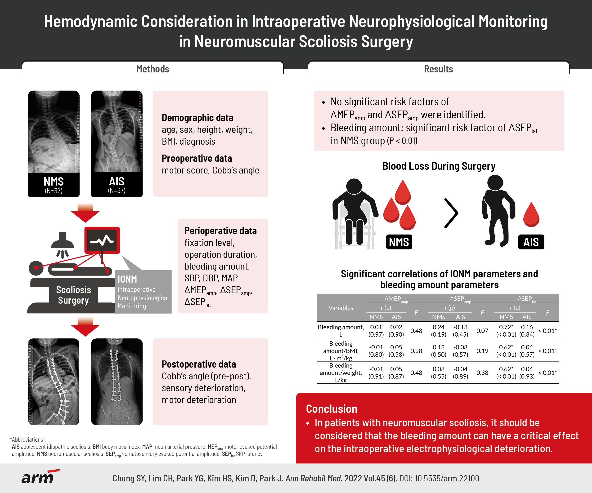
Simple regression analyses were used to analyze each risk factor affecting the IONM parameters (ΔMEPamp, ΔSEPamp, and ΔSEPlat) in the NMS and AIS groups (Table 2). No significant risk factors of ΔMEPamp and ΔSEPamp were identified. Bleeding amount (p<0.01) was analyzed as a significant risk factor of ΔSEPlat in NMS group, but the other variables did not significantly contribute to the ΔSEPlat in either the NMS or AIS group.
Bleeding amount and IONM parameters
As listed in Table 1, the bleeding amount showed significant differences between the NMS and AIS groups. As significant differences in preoperative body weight and BMI were found between the two groups, “bleeding amount/BMI” and “bleeding amount/weight” were used to correct the bleeding amount for body weight or BMI (Table 3). The NMS/AIS ratios of the bleeding-related parameters were as follows, with statistically significant intergroup differences: bleeding amount (1.56, p<0.01), bleeding amount/BMI (2.13, p<0.01), and bleeding amount/weight (2.62, p<0.01). When the bleeding amount was corrected by BMI or body weight, the difference in bleeding amount ratio between the two groups was found to be greater.
We confirmed through correlation analyses whether a significant correlation existed between the IONM and bleeding-related parameters, and the differences between the two groups were confirmed through Fisher transformation (Table 4). In the NMS group, positive correlations were found between ΔSEPlat and all the bleeding-related parameters as follows: bleeding amount (r=0.72, p<0.01), bleeding amount/BMI (r=0.62, p<0.01), and bleeding amount/weight (r=0.62, p<0.01), where ΔMEPamp and ΔSEPamp did not show any correlation with any of the three bleeding-related parameters. In the AIS group, no correlation was revealed between the IONM and bleeding-related parameters.
Correlation between bleeding amount and BP
All three bleeding-related parameters significantly correlated with MAPmin, SBPmin, and DBPmin in the NMS group, where they did not correlate with MAPmin, SBPmin, and DBPmin in the AIS group (Table 5).
DISCUSSION
The bleeding amount was a significant contributing factor to SEP latency prolongation in NMS as shown in Tables 2 and 4. After excessive bleeding, ischemic condition may result from hypotension or hypoxia. To our knowledge, no previous studies have examined the direct relationship between bleeding amount and IONM parameters in scoliosis surgery. However, previous studies confirmed that decreased blood flow, BP reduction, and following hypoxic conditions contributed to the deterioration of SEP amplitude and latency [14,15]. In a posttraumatic animal model, Fehlings et al. [16] reported that both MEPs and SEPs were significantly related to posttraumatic blood flow, which contributes to the severity of spinal cord injury. Banoub et al. [17] showed that when MAP decreased more than a certain value, the SEP amplitude decreased without a change in latency. According to McPherson et al. [18], severe hypoxia led to decreased SEP amplitude and increased SEP latency in canis experiment.
Before the demonstration of the effect of ischemic condition on the SEPs along with the posterior column, ischemia had already been revealed to prolong the latency and reduce the amplitude of the peripheral nerves in conduction study [19]. Temporal dispersion was reported to follow after the anoxia in peripheral nerves, and with the previous evidence that the reduction in a single-fiber spike amplitude does not exceed 16%, the major cause of the amplitude reduction in peripheral nerves is explained by temporal dispersion [19-21]. As SEPs are used to evaluate the electrophysiological state along the extension of the peripheral sensory nerve to the central nervous system, the decreased amplitude due to bleeding-related ischemic condition would also result from temporal dispersion, not from conduction block. Especially, in cases where the spinal cord is not directly exposed as in scoliosis surgery, the damage of the spinal cord within the spinal canal is suspected by mechanical correction of the curvature rather than direct iatrogenic injury.
Clinicians have no choice but to question in what order the electrophysiologic parameters deteriorate when the ischemic state of the patient prolongs during surgery, and related animal experiments have been conducted. Reuter et al. [22] reported that SEP disappeared ahead of the loss of MEP waveform as hypoxia continued in a canis model. Lesnick et al. [23] reported that as global ischemia worsens, latency prolongation precedes the amplitude reduction of SEPs in a felis model. In turn, in scoliosis surgery, the mechanism of the deterioration of the IONM parameters in ischemic conditions is not conduction block, which can result from direct neural injury, but temporal dispersion from the gradual slowing of conduction velocity. This means that for the SEP amplitude to decrease to a certain level, latency must also be delayed to a certain level. From a preventive medicine point of view, even with a neurophysiological state of confirmed prolonged SEP latencies, if hemodynamic conditions are stably maintained by transfusion, fluid supplementation, or use of vasopressors, it may not lead to a significant reduction in SEP amplitude. There is a previous experiment that the electrophysiologically compromised condition of spinal cord can be restored by reperfusion. According to a rabbit experimental study by Ji et al. [24], when reperfusion was performed in the spinal cord for >20 minutes in ischemic injury models, the latency gradually returned to normal levels, while the amplitude could not recover, which means that axonal degeneration has occurred while myelin is preserved. Thus, when IONM professionals face SEP latency prolongation without amplitude decline, they have to pay attention more to the bleeding amount and the following BP and maintain close communication with the anesthesiologist and the surgeons to stabilize hemodynamic conditions. A significant decrease in SEP amplitude after an excessive bleeding event would be of course more urgent situation that can lead to postoperative neurological deterioration. Schwartz et al. [25] reported that in three patients with weakness after scoliosis surgery due to hypotension, SEP amplitude reduction was lagged by 5 minutes from MEP amplitude reduction. However, they did not count SEP latency, and there is no description of the relationship of hemodynamic changes with SEPs or MEPs in patients without postoperative neurological deterioration. From this point of view, our study has strength in that it provides information on subtle early changes in the parameters before postoperative neurological deterioration occurs rather than discussing about the neurophysiological relationship after the event occurs.
As the anesthesiologist and the surgical nurses continuously check the suctioned fluid and calculate the estimated blood loss, a precise modulation of the vital signs is continued through the whole surgical procedure. However, in this study only the NMS group showed significant correlation of the bleeding amount to the parameters of blood pressure: MAPmin, SBPmin, and DBPmin (Table 5). This implies that the rate of supplementation for blood loss may not catch up with the rate of BP reduction in the NMS group, where the supplements for blood loss in the AIS group were sufficient to maintain the BP. In addition, the NMS/AIS ratios of the bleeding-related parameters were higher in the order of bleeding amount/weight, bleeding amount/BMI, and bleeding amount (Table 3). We can infer that bleeding-induced hypovolemia compared with body weight is a more important factor of the BP decrement in the NMS group. The bleeding amount has been reported to be greater in scoliosis surgery for NMS than in that for AIS [26,27]. In addition, as seen in Table 1, the bleeding amount was greater in NMS than in AIS. This may be due to the different hemodynamic physiology between the two groups. Previously, hemodynamic instability in neuromuscular diseases was observed. BP was lower in patients with Duchenne muscular dystrophy (DMD), a representative neuromuscular disease, than in healthy patients, and this was thought to be due to decreased cardiac function [28,29]. In addition, it is reported that an increase in prothrombin time and depletion of factor VII is more common in NMS than AIS, which can lead to an increase in the bleeding amount [30].
In our study, although the bleeding amount was greater in the NMS group, none of the patients showed postoperative neurological deterioration after surgery in both the NMS and AIS groups, so that it was difficult to confirm the relationship between blood loss and postoperative neurological prognosis. However, some studies have demonstrated that the bleeding amount during scoliosis surgery is closely related to postoperative prognosis. Carreon et al. [31] showed that the greater the bleeding amount, the higher the frequency of complications after AIS surgery. Jia et al. [32] reported that NMS patients receive more transfusions due to intraoperative excessive bleeding, increasing their risk of exposure to transfusion side effects. Therefore, excessive bleeding during scoliosis surgery should be prevented, and blood loss should be compensated quickly and properly. Through proper transfusion or vasopressor use, postoperative neurological deterioration due to hypotension or hypoxia could be prevented. In this course, the SEP may provide early clues of the ischemia-related neurophysiological deterioration, enabling rapid response to hemodynamic instability.
This study has several advantages. First, it may provide evidence of the relationship between bleeding amount and electroneurophysiological changes in scoliosis surgery. Second, it was suggested that hemodynamic and electrophysiological changes to bleeding appear differently according to pathological characteristics in NMS and AIS. As real-time monitoring of electrophysiological status, rather than the bleeding amount itself has a more direct correlation with prognosis, it is suggested that the hemodynamic supplementation should be prompted after massive bleeding while confirming the IONM parameter is restored [33]. Especially, SEP latency is suggested as an important indicator which reflect the ischemic condition without direct axonal damage. This study has a limitation in that there was no case of postoperative neurological deterioration, so it might be not possible to review cases with severe hemodynamic instability. On the other hand, however, it may be a study that provides evidence that the parameters of SEP respond to hemodynamic changes ahead of changes in MEP occurs. Previous studies have been emphasized that it is important to monitor the SEPs together with the MEPs, even in studies analyzing the reliability of IONM. Nevertheless, on the basis that SEP does not anatomically reflect the physiology of the motor tract, the monitoring of SEP is still underestimated by some surgeons and examiners. This study will be useful in convincing those who merely rely on MEPs that it is much better to monitor SEPs in conjunction with MEPs. If a large sample study, including cases with more diverse neurological prognosis under IONM could be performed, it would be helpful to understand the correlation between hemodynamics and electroneurophysiology in scoliosis surgery in more detail.
Bleeding amount is an important factor in prolonging SEP latency during NMS surgery. The effect of bleeding on neurophysiological changes is stronger in NMS than in AIS. Based on the result that the SEP latency is more vulnerable than SEP or MEP amplitude in ischemic conditions, it is suggested that SEP latency could be an early index for the ischemic injury occurs without direct axonal injury during scoliosis surgery. Therefore, when SEP deterioration occurs with massive bleeding, it is important for IONM specialists, surgeons, and anesthesiologists to communicate intensively to stabilize the hemodynamic state until SEP latency is restored.