Optimal Placement of Needle Electromyography in Extensor Indicis: A Cadaveric Study
Article information
Abstract
Objective
To identify the center of extensor indicis (EI) muscle through cadaver dissection and compare the accuracy of different techniques for needle electromyography (EMG) electrode insertion.
Methods
Eighteen upper limbs of 10 adult cadavers were dissected. The center of trigonal EI muscle was defined as the point where the three medians of the triangle intersect. Three different needle electrode insertion techniques were introduced: M1, 2.5 cm above the lower border of ulnar styloid process (USP), lateral aspect of the ulna; M2, 2 finger breadths (FB) proximal to USP, lateral aspect of the ulna; and M3, distal fourth of the forearm, lateral aspect of the ulna. The distance from USP to the center (X) parallel to the line between radial head to USP, and from medial border of ulna to the center (Y) were measured. The distances between 3 different points (M1– M3) and the center were measured (marked as D1, D2, and D3, respectively).
Results
The median value of X was 48.3 mm and that of Y was 7.2 mm. The median values of D1, D2 and D3 were 23.3 mm, 13.3 mm and 9.0 mm, respectively.
Conclusion
The center of EI muscle is located approximately 4.8 cm proximal to USP level and 7.2 mm lateral to the medial border of the ulna. Among the three methods, the technique placing the needle electrode at distal fourth of the forearm and lateral to the radial side of the ulna bone (M3) is the most accurate and closest to the center of the EI muscle.
INTRODUCTION
The extensor indicis (EI) muscle, which is the narrow and trigonal skeletal muscle supplied by the posterior interosseous nerve [1], originates from the posterior surface of the ulnar and the adjacent interosseous membrane. Its tendon is inserted into the index finger via the extensor expansion [2]. It is commonly used to record the compound muscle action potential of the radial nerve. Needle electromyography (EMG) of the EI muscle also facilitates the diagnosis of C8 radiculopathy and radial nerve lesion. This muscle is also one of the target muscles for botulinum toxin injection in patients with focal hand dystonia [3]. However, due to its small size, oblique pathway, and covering with a superficial layer of muscles in the posterior compartment of the forearm [4], accurate placement of the recording electrode in the EI muscle is a challenge.
Several techniques for needle EMG of the EI muscle have been proposed. Chu-Andrews and Johnson [5] recommended insertion of the needle 2.5 cm proximal from the lower border of styloid process of ulnar, in line with the lateral aspect of the ulna with forearm pronated. Perotto and Delagi [6] preferred to insert the needle at 2 finger breadths (FB) proximal to the ulnar styloid, just radial to ulnar at a depth of one-half inch. Lee and Delisa [7] suggested needle insertion in the distal fourth of the forearm lateral to the radial side of the ulnar between the extensor digitorum and extensor carpi ulnaris tendons. We were motivated to compare the accuracy of the different needle insertion methods. The purpose of this study was to identify the center of the EI muscle through cadaver dissection and compare the accuracy of different methods of needle EMG electrode insertion.
MATERIALS AND METHODS
Eighteen upper limbs of 10 adult cadavers were dissected. We missed two upper limbs because they were already dissected in the other study. None of the recorded causes of death affected the results of this study. The skin and subcutaneous tissue were dissected first. After the superficial layer of muscles in the posterior compartment of the muscle was dissected, EI was exposed. The most proximal part of EI, where the muscle is attached to the ulna, just distal to extensor pollicis longus muscle, was designated as the proximal origin (O2) and the most distal site of the muscle anchored to the ulna was marked as the distal origin (O1). The musculotendinous junction (MT) was the distal and middle point where the muscle changes to the tendinous portion. As shown in Fig. 1, the center of EI (C) was defined as the centroid of the triangle formed by these three points (O1, O2, and MT). The center of EI (C) was defined as the centroid because of the low possibility of missing the triangular muscle in conditions targeting the centroid. The needle insertion was carried out according to three different methods [5-7]: 2.5 cm above the lower border of the ulnar styloid process (USP), lateral aspect of the ulna (M1); 2 FB (approximately 30 mm in this study) proximal to USP, lateral aspect of the ulna (M2); and distal fourth of the forearm, lateral aspect of the ulna (M3).

Schematic diagram of center (C) of extensor indicis (EI) muscle (left forearm pronated with dorsal view). (A) The triangle formed by three points (O1, O2, and MT) in EI muscle. (B) The center of EI (C) was defined as the centroid of the triangle. O1, distal origin of EI muscle; O2, proximal origin of EI muscle; MT, musculotendinous junction of EI; USP, the lower border of ulnar styloid process.
As shown in the Fig. 2, the distance from USP to C (C_X) parallel to the line between radial head to USP, and from medial border of ulna to C (C_Y) were measured. The forearm length (FL) was measured from the radial head to USP. The forearm width (FW) at the level of C was measured. The ratios of C_X to FL and C_Y to FW were also calculated as a percentage.

Parameters measured in the cadaver (left forearm pronated with dorsal view). A, angle between ulna and the line connecting the proximal origin of extensor indicis (EI) muscle and C; MT, musculotendinous junction of EI muscle; M1, 2.5 cm above the lower border of USP lateral aspect of the ulnar bone; M2, 2 finger breadths proximal to USP, lateral aspect of the ulna; M3, distal fourth of the forearm, lateral aspect of the ulna; O1, distal origin of EI muscle; O2, proximal origin of EI muscle; C, center of EI muscle; USP, tip of ulnar styloid process; C_X, distance between USP and C; C_Y, distance from medial border of ulnar bone to C.
The distance between three different points (M1–M3) and C were measured (D1, D2, and D3, respectively) to assess and compare the accuracy of different methods. The angle (A) between ulna and the line connecting the proximal origin of EI and C was measured (Fig. 2).
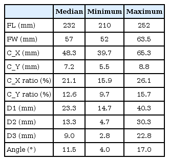
The median and range (minimum-maximum) of each parameter were recorded in Table 1.
RESULTS
The median distance from radial head to USP (FL) was 232 mm, and the FW was 57 mm. The median distance from USP to C parallel to the line between radial head to USP (C_X) was 48.3 mm and the percentage of C_X to total FL was 21.1%, which suggests that the center (C) of EI was approximately distal 20.0% of total FL. The median distance from medial border of ulna to C (C_Y) was 7.2 mm and the percentage of C_Y to forearm width (FW) was 12.6%.
The median distances between three different points (M1–M3) and C (D1, D2, and D3, respectively) were 23.3 mm, 13.3 mm, and 9.0 mm, respectively, which suggests that the point distal fourth of the forearm and lateral to the radial side of the ulnar bone, is close to the mid-point of EI muscle. The median angle between ulna and the line connecting the proximal origin of EI was 11.5°.
DISCUSSION
Our study demonstrated that the center of EI muscle was located approximately 5 cm proximal to the USP level (distal 20% of FL) and 0.7 cm lateral to the medial border of ulna (12.6% of FW). This location was consistent with the needle insertion site described by Lee and DeLisa [7], in which the distance from the center of EI muscle was less than 1 cm. The method described by Perotto and Delagi [6] was less precise due probably to the use of examiner’s finger width, which varies among examiners. The method by Chu-Andrews and Johnson [5] based on an absolute fixed distance (2.5 cm) may also be less reliable because of varying length of individual’ forearms.
The varied needle insertion methods across different studies suggest the difficulty of accurate localization of the EI muscle. In the previous study by Karvelas et al. [8], the accuracy of needle placement in each of the four selected muscles was investigated: EI, pronator teres, peroneus longus, and soleus in live subjects. They confirmed the needle localization by ultrasound. The overall accuracy of needle electrode localization was 68.8% for all the muscles tested. Localization of the EI muscle was the least accurate, demonstrating accurate placement by 20% to 42.9%. In a cadaver study, Goodmurphy et al. [9] demonstrated that smaller and deeper muscles were harder to hit than larger and superficial muscles. In the upper limb, the serratus anterior, flexor carpi ulnaris, flexor carpi radialis, flexor pollicis longus, pronator teres, and EI were harder to hit.
Inaccurate needle insertion increases the possibility of hitting an incorrect muscle, compromising the diagnostic utility and leading to misdiagnosis [10]. Insertion of the needle electrode proximal to the EI muscle may lead to location in the extensor pollicis longus muscle [7]. When the needle electrode is placed too distally, it may be positioned in the tendon portion of EI muscle. Our results are expected to facilitate the estimation of two-dimensional but not three-dimensional location. The needle electrode was inserted too deeply and the pronator quadratus was sampled [9]. If the needle electrode was superficially located, it may be placed in the extensor carpi ulnaris or extensor digiti minimi muscle [11]. In these cases, we need to discriminate motor unit action potentials of the EI muscle from those of other muscles. Further study is needed to determine the accurate depth of the EI muscle.
The anatomic motor point has been defined as the entry point of the motor nerve branch into the epimysium of the muscle belly [12]. The active recording electrode in nerve conduction study should be placed over the motor point. In our study, we did not identify the motor point but estimated the center of the EI muscle. A previous anatomical study reported that the motor points for the EI muscle were located in the middle third of the muscle belly [12]. Therefore, it is likely that the center of EI muscle is located in the territory of motor points.
The blockade of the EI muscle activity in patients with focal hand dystonia with motor point block may be effective using botulinum toxin. However, the toxin may be injected into the wrong muscle group causing paralysis of unintended muscles or requiring higher doses [13]. Animal studies demonstrated that botulinum toxin diffused as far as 4.5 cm from the injection site and crossed fascial planes to adjacent non-injected muscles [13]. Thus, the botulinum toxin injection around the motor point estimated in our results can increase efficacy and reduce the unexpected effect.
There are several limitations in this study. First, it is impossible to present the mean and standard deviation of the results in this study due to the small number of cadavers dissected. Further studies are needed to obtain statistically robust results. Second, variation in height and forearm length of cadaver and width of FB between different testers was not based on different racial groups. This variation affects the location of M1–M3 and results of D1–D3.
In conclusion, the center of EI muscle is located distal fifth of the FL and just lateral to the ulna, and is consistent with the needle insertion technique described by Lee and DeLisa [7]. The results are expected to provide valuable insight into nerve conduction study and needle EMG, and facilitate the determination of injection site of botulinum toxin.
Notes
No potential conflict of interest relevant to this article was reported.

