• KARM
  • Contact us
  • E-Submission
ABOUT
ARTICLE TYPES
BROWSE ARTICLES
AUTHOR INFORMATION

Articles

Original Article

The Effect of Extracorporeal Shock Wave Therapy on Myofascial Pain Syndrome

Jong Hyun Jeon, M.D., Yun Jae Jung, M.D., Ju Youn Lee, M.D., Ji Soo Choi, M.D., Jeong Hyeon Mun, M.D., Won Yong Park, M.D., Cheong Hoon Seo, M.D., Ki Un Jang, M.D., Ph.D
Annals of Rehabilitation Medicine 2012;36(5):665-674.
Published online: October 31, 2012

Department of Rehabilitation Medicine, Hallym University Burn Institute, Hallym University College of Medicine, Seoul 150-719, Korea.

Corresponding author: Ki Un Jang. Department of Rehabilitation Medicine, Hallym University Burn Institute, Hallym University College of Medicine, Yeongdeungpo-dong 2-ga, Yeongdeungpo-gu, Seoul 150-719, Korea. Tel: +82-2-2639-5730, Fax: +82-2-2633-7571, jangkiun@hanmail.net
• Received: March 26, 2012   • Accepted: July 13, 2012

Copyright © 2012 by Korean Academy of Rehabilitation Medicine

This is an open-access article distributed under the terms of the Creative Commons Attribution Non-Commercial License (http://creativecommons.org/licenses/by-nc/3.0) which permits unrestricted noncommercial use, distribution, and reproduction in any medium, provided the original work is properly cited.

  • 6,983 Views
  • 160 Download
  • 58 Crossref
  • 61 Scopus
prev next
  • Objective
    To investigate the effect of extracorporeal shock wave therapy (ESWT) on myofascial pain syndrome (MPS).
  • Method
    Thirty patients with MPS in trapezius muscle were randomly divided into two groups, ESWT group (n=15), and trigger point injections (TPI)+transcutaneous electrical nerve stimulation (TENS) group (n=15). For a total of 3 weeks, ESWT was undertaken with 1,500 pulse each time at one week interval totaling 4,500 pulse, TPI for once a week totaling three times and TENS for five times a week totaling three weeks.
  • Results
    The changes in pain threshold (lb/cm2) showed the values of 6.86±1.35 before first therapy, 11.43±0.27 after first therapy, and 12.57±0.72 after third therapy, while TPI+TENS group showed the values of 6.20±1.92 before first therapy, 8.80±0.48 after first therapy, and 9.60±2.19 after third therapy, and the changes between the groups were significantly different (p=0.045). The changes in visual analog scale were estimated to be 6.86±0.90 before first therapy, 2.86±0.90 after first therapy, and 1.86±0.69 after third therapy in case of ESWT group, whereas the figures were estimated to be 7.20±1.30 before first therapy, 4.60±0.55 after first therapy, and 2.80±0.84 after third therapy in case of TPI+TENS group, and the changes between the groups were significantly different (p=0.010). The changes in McGill pain questionnaire (p=0.816) and pain rating scale (p=0.644) between the groups were not significantly different. The changes in neck ROM were also not significantly different between the groups (p>0.05).
  • Conclusion
    The ESWT in patients with MPS in trapezius muscle are as effective as TPI and TENS for the purpose of pain relief and improving cervical range of motion.
Myofascial pain syndrome is known to be a clinically common syndrome with characteristics including localized muscle tenderness, typical referred pain, palpable intramuscular taut band and muscular twitching response, etc., and induction of pain due to the myofascial trigger points (MTrPs) is defined as hyperirritable spot of taut band.
Myofascial pain syndrome is a problem frequently experienced in clinical setting that accounts for the largest proportion of the diseases of musculoskeletal system occurring in cervical and lumbar vertebral portion, and shoulder, and is a common cause that induces disability due to pain. The basic diagnostic method of myofascial pain syndrome is determined by physical examination such as localized muscle tenderness, typical referred pain, palpable intramuscular taut band, muscular twitching response along with subjective symptoms of the patients. In addition, method of assessment of pain threshold using algometry can be helpful in diagnosing myofascial pain syndrome. As the methods of treatment for myofascial pain syndrome, stretching, dry-needling and trigger point injection using diverse range of drugs are used along with generally practiced physical therapy.1
Recently, application of Extracorporeal Shock Wave Therapy (ESWT) has been expanded as one of the treatment methods for the diseases of musculoskeletal system. Although there have been reports about the effectiveness of ESWT mainly on epicondylitis, plantar fasciitis, chronic pelvic pain, chronic painful heel syndrome, lymphedema, burn wound, pressure ulcer and calcific tendinitis, its effectiveness on treatment of myofascial pain syndrome has not been reported. Accordingly, this study aims to examine the effectiveness and clinical usefulness of ESWT by comparing the ESWT implemented group with the trigger point injection (TPI) and transcutaneous electrical nerve stimulation (TENS) implemented group with myofascial pain syndrome patients as the subjects.
The authors selected 30 patients who were diagnosed with myofascial pain syndrome in the trapezius muscle as a result of physical examination with pain in the posterior neck and shoulder areas as the chief complaint among the hospitalized patients at our hospital from January to December 2011 as the subjects of this study. This study is a prospective randomized experimental and control group research with the subjects divided into 2 groups, namely, the experimental group with 15 subjects who underwent ESWT, and the control group with 15 subjects who underwent TPI and TENS. For ESWT treatment, subjects were given total of 1,500 shock waves for each treatment at the rate of 240 waves, each with low energy flux density (EFD) of 0.10 mJ/mm2 per minute by using EvoTron RFL0300 of SwiTech Medical AG 3 times with a week's interval between the treatments (total of 4,500 shock waves). Treatment was given by focusing on the area at which muscular twitching response or referred pain should be induced by appropriately adjusting the location of the localized probe. Total of 3 TPI treatments with a week's interval between the treatment and total of 5 TENS treatments were given 5 times in a week with a duration of 20 minutes a day. Both the groups underwent treatment for total of 3 weeks each. Visual Analogue Scale (VAS), McGill pain questionnaire, Pain Rating Scale (PRS), neck range of motion (curvature, extension, rotation, lateral bending) and BASELINE® (Hoggan Health Industries Inc., West Jordan, USA) were used to confirm the changes in the pain threshold level (lb/cm2) prior to the first treatment (preliminary assessment), 1 week after the first treatment (interim assessment) and 1 week after the third treatment (final assessment) as the assessment method for assessing the effectiveness of treatment. At the time of final assessment, the results were categorized on the basis of assessment of Roles and Maudsley score and whether MTrPs was induced and referred pain was extinct, 'Excellent' if both have been extinct, 'Good' if only one of the 2 has been extinct and 'Poor' if both have not been extinct for additional execution of final response assessment.
Collected data were analyzed by using SPSS 19.0 program (SPSS Inc., Chicago, USA). Fisher's Exact test and Mann-Whitney test were carried out for homogeneity test on the 2 groups. Since the result of review of the normality assumption on the preliminary assessment tools of the 2 groups was satisfactory, Independent Samples t-Test was carried out to verify the preliminary homogeneity, and Repeated Measure ANOVA was carried out for examination of the effectiveness of the results of preliminary assessment, and assessment following the 1st and the 3rd treatment. Prior to execution of Repeated Measure ANOVA, Sphericity test of Mauchly for measurement of homoscedasticity and compound symmetry was performed. In the case of satisfaction of the sphericity assumption (p>0.05), verification of time effect (Time) and reciprocal action effect (Group×Time) were analyzed by using sphericity assumption statistics of Univariate test. If sphericity assumption was not satisfied (p<0.05), statistics of Wilks' Lambda in Multivariate test was used. Moreover, Fisher's exact test of small specimen was carried out for verification of the results of Roles and Maudsley score and final response assessment. Lastly, in order to find out if there are differences in the results of treatment between the 2 groups due to gender difference, gender difference was confirmed by using Independent Samples t-Test in the final assessment.
Demographic and clinical characteristics of the patients
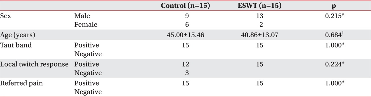
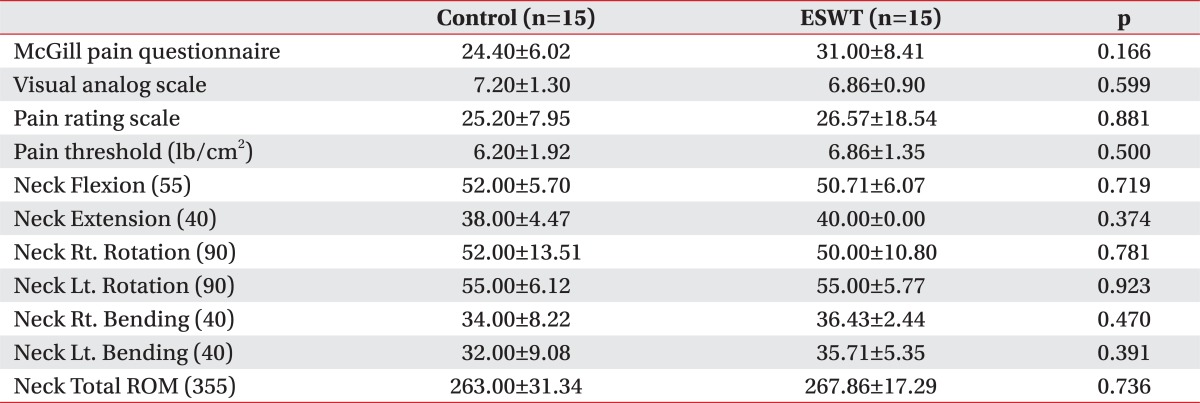
A total of 30 patients comprised 22 males and 8 females. Of the 15 subjects in the experimental group, 13 were male and 2 were female, while the control group composed of 9 males and 6 females. The average age was 40.86±13.07 years for the experimental group and 45.00±15.46 years for the control group. As a result of the physical examination, taut band and referred pain were observed in both, the experimental and the control group. Localized twitching response was observed in all the 15 subjects of experimental group while 12 of the 15 subjects of the control group displayed the response. As a result of Fisher's Exact test and Mann-Whitney test carried out to examine the preliminary homogeneity between the 2 groups, both the groups were found to be homogeneous as there was no significant difference (p>0.05) (Table 1). As a result of Independent Samples t-Test carried out to examine the preliminary homogeneity of the preliminary assessment tools, preliminary measurement tools were found to be homogeneous as there was no significant difference (p>0.05) (Table 2).
The change in pain threshold
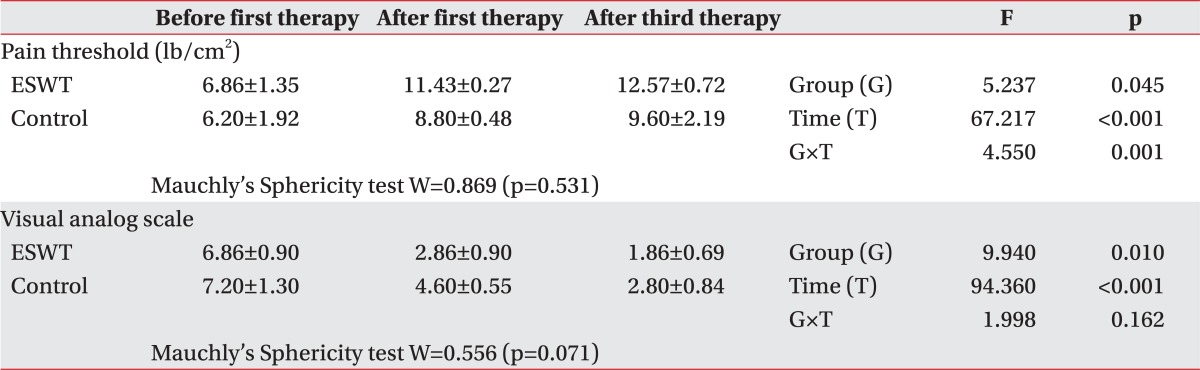
As a result of Repeated Measure ANOVA on pain threshold, it was found that there was significant difference in accordance with groups (p=0.045<0.05), time effect (p<0.001) and reciprocal action effect (p=0.001<0.01). As a result of contrast test on time, a significant difference was observed prior to commencement, following the 1st and the 3rd treatment, illustrating that the pain threshold began to increase after the 1st treatment. Effect of the group was found to be significant, resulting in higher pain threshold in the ESWT group in comparison to the TPI and TENS group. As a result of contrast test on the reciprocal action, there was significant difference prior to commencement, following the 1st and the 3rd treatment, illustrating that the treatment effect in the ESWT group was greater than the TPI and TENS group since the 1st treatment (Table 3).
The changes in visual analog scale
As a result of sphericity on VAS, it was found that sphericity assumption was satisfied (p=0.071>0.05), and it was also found that although there was significant difference in VAS in accordance with groups (p=0.010<0.05) and time effect (p<0.001), there was no reciprocal action effect (p=0.162>0.05). As a result of contrast test on time, there was significant difference in VAS prior to commencement, following the 1st and the 3rd treatment. Therefore, a significant reduction in VAS was noted following the treatment. Although there was significant difference in VAS between the groups, it was found that there was no reciprocal action effect. This signifies that treatment using ESWT is more effective than the treatment using TPI and TENS since the VAS of the ESWT group was found to be lower than that of the TPI and TENS group (Table 3).
The changes in McGill pain questionnaire
Sphericity assumption was satisfied as a result of Repeated Measure ANOVA on McGillpain questionnaire (p=0.666>0.05). Although there was no significant difference between the groups (p=0.816>0.05), time effect (p<0.001) and reciprocal action effect (p=0.031<0.05) was observed. As a result of contrast test on time, it was found that there was reduction following the 1st and the 3rd treatment in comparison to prior to commencement, thus illustrating that McGill pain questionnaire was significantly reduced as the treatment was carried out. There was significant difference in the results of contrast test on reciprocal action prior to commencement and following the 3rd treatment. This signifies that although the McGill pain questionnaire prior to commencement was found to be higher in the ESWT group, it was lower in the ESWT group following the 3rd treatment and that treatment using ESWT had greater effectiveness after the 3rd treatment (Table 4).
The changes in pain rating scale
Sphericity assumption was found to be unsatisfactory based on the result of sphericity on PRS (p<0.001). Based on the Repeated Measure ANOVA carried out by Wilks' Lambda, it was found that there was no significant difference between the groups (p=0.644>0.05) but also no reciprocal action effect (p=0.806>0.05), and only the time (p=0.004<0.01) effect was found to be significant. As a result of contrast test on time, significant difference prior to commencement and following the 1st and the 3rd treatment was observed, illustrating that PRS was reduced significantly since the 1st treatment (Table 4).
The changes in neck range of motion
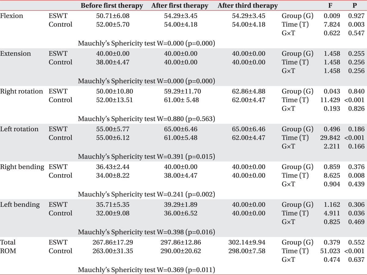
As a result of the Repeated Measure ANOVA carried out on neck range of motion, there was no significant difference between the groups (p>0.05) but also no reciprocal action effect (p>0.05) in all the category, and, in all the categories excluding extension, time (p<0.05) effect was found to be significant. Therefore, it was found that there was effectiveness of treatment on both the ESWT group and TPI and TENS group in terms of neck range of motion (Table 5).
Final response assessment
As a result of measurement of Roles and Maudsley score, experimental group had 6 subjects with rating of excellent and 9 subject with rating of good among total of 15 subjects while control group had 3 subjects with rating of excellent, 9 subjects with rating of good and 3 subjects with rating of acceptable among the total of 15 subjects. There was no significant difference between the groups (p=0.233>0.05).
As a result of measurement in the final response assessment, experimental group had 10 subjects with rating of excellent and 5 subjects with rating of good among total of 15 subjects while control group had 6 subjects with rating of excellent and 9 subjects with rating of good among total of 15 subjects. There was no significant difference between the groups (p=0.272>0.05).
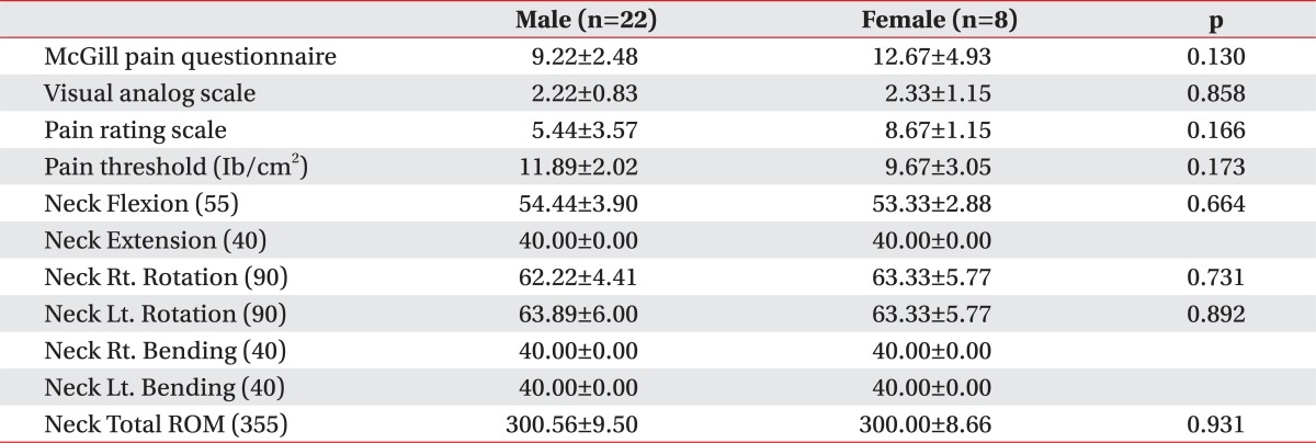
Gender difference on the final assessment
As a result of confirmation of the differences between the gender in the final assessment in order to determine the differences in the results of treatment in the 2 groups due to gender ratio, no significant gender difference (p>0.05) was observed (Table 6).
The ESWT was first applied in Germany in 1996 and following 5 diseases to which ESWT can be applied clinically were proposed: (1) calcific tendonitis, (2) lateral epicondylitis, (3) medial epicondylitis, (4) plantar fasciitis, and (5) pseudoarthrosis.2 Typical characteristic of the shock wave generated in ESWT is that pressure reaches 100 MPa or 1,000 times that of atmospheric pressure during short period of several nanoseconds, which then gradually falls to the normal level prior to turning into negative pressure. The negative pressure equivalent to approximately 10% of the size of the maximum pressure generated is the characteristic of the shock wave and cavitational effect is generated through this characteristic feature. Shock wave used for medical purposes includes extracorporeal shock wave lithotripsy (ESWL) and ESWT, which are used by focusing the shock wave generated outside the body on the target area in patients with urolithiasis or orthopedic diseases.3
The ESWT equipment currently used in clinical setting is divided into radial and focused methods in accordance with the manner in which the shock wave reaches the target. There are 3 methods of generating focused-type shock wave, namely, piezoelectric, electromagnetic and electrohydraulic methods. Shock wave is generated by using the principle in which electricity is converted into shock wave through quick physical movement of electricity in a liquid medium. Each of the equipment uses charged condenser with different voltage from each other, and conversion into shock wave is achieved as electricity becomes rapidly discharged in acoustic transducer.3 ESWT can be classified in accordance with the level of energy flux density (EFD) into as low energy with EFD range of 0.08-0.27 mJ/mm2, intermediate energy with EFD range of 0.28-0.59 mJ/mm2, and high energy with EFD of more than 0.6 mJ/mm2. In addition, although another standard for classification into low energy for EFD of less than 0.12 mJ/mm2 and high energy for EFD of more than 0.12 mJ/mm2 has been presented,4 there is no definitively unified standard yet. Electrohydraulic format of low energy flux density used in this study in which shock wave is generated in the typical format of pressurized wavelength that rapidly increases within extremely short period of time and then decreases gradually is known to have little side effects and excellent effectiveness as per the results of laboratory research,3,5 and low energy extracorporeal shock wave therapy, in comparison to treatment using high energy, requires no localized anesthesia at the time of treatment and is known to prevent the interference of recovery of the damaged tissues and rupture of tissues that could occur with high energy extracorporeal shock wave therapy.3,6 In addition, by using low energy, it is possible to prevent the phenomenon of lowering of the level of adaptation of the patients or giving up of the treatment in the middle by the patients, due to the pain caused by the treatment itself that occurs when high energy shock wave is used. However, histological reaction on extracorporeal shock wave is known to be dose-dependent on the total energy delivered (Total Effectiveness Energy=EFD (mJ/mm2)×mm2×number) (mJ)3,7 and, low-energy treatment has the disadvantage of having to perform treatment several times in order to maintain the total energy delivered to the area to be treated at the level similar to that of high energy extracorporeal shock wave.
Until now, there is no guideline that clearly defines the number of and interval for execution of ESWT. In the study by Jung et al.,8 in which low energy shock wave treatment for chronic epicondylitis of elbow joint was carried out, approximately 2,000 shock waves with low EFD range of 0.06-0.12 mJ/mm2 were delivered in accordance with the pain tolerance of the patient during each treatment for total of 3 times with a week's interval between treatments, and, in the study by Kim et al.9 on the effectiveness of extracorporeal shock wave therapy on plantar fasciitis patients, 1,200 shock waves with low EFD of 0.24 mJ/mm2 were delivered for total of 3 times with a week's interval between the treatments. In this study, subjects were given total of 1,500 shock waves for each treatment at the rate of 240 waves, each with low energy flux density (EFD) of 0.10 mJ/mm2 per minute by using for total of 3 times with interval of a week between the treatments (total of 4,500 shock waves). When shock wave was applied to the accurate area at the time of treatment, it was possible to confirm that induction of pain and referred pain as well as muscular twitching response occurred in the entire experimental group.
In general, it is known that the effect of extracorporeal shock wave in living tissues induces characteristic changes within the cells due to conversion of the mechanical signal into biochemical or molecular biologic signal (Mechanotransduction).10 According to the available knowledge, although the mechanisms of ESWT are not clear, several hypothesis have been discussed on the principles of cellular and molecular biological effects. According to Zimmermann et al.,10 ESWT improves blood circulation in capillary blood vessels and reduces the tension and stiffness of muscles along with the reduction in pain by inducing interference of flow of excessive stimulation of nociceptors and stimulation of nerves. In addition, the study carried out by Hausdorf et al.11,12 states that ESWT reduces pain in the tissues of musculoskeletal system through selective destruction of nonmyelinated fibers and is effective in reducing the level of substance P in the target tissue as well as reducing the synthesis of substance P in dorsal root ganglia. According to Wang et al.,13 ESWT is known to be effective in acceleration of perfusion in the ischemic tissues (myocardium and skin flap) and stimulation of generation of new blood vessels, and according to Davis et al.,14 it is effective in recovering the areas of ischemic skin flap and increasing the perfusion of tissues through reduction effect on inflammation. According to De Sanctis et al.,15 ESWT is effective in leading to improvement of circulation in capillary blood vessels in the chronic ischemic areas of lower limbs when it is performed on people and according to Fukumoto et al.,16 it is effective in improving the perfusion of myocardium in patients with severe diseases of coronary artery. As per the study by Sparsa et al.,17 ESWT has been reported to reduce pain and calcification and accelerate re-epithelization in CREST syndrome and chronic leg ulcer.
In a study by Muller-Ehrenberg and Licht,18 focused-type extracorporeal shock wave therapy was carried out on 30 patients with myofascial pain syndrome, pain and typical referred pain were induced in 95% of the subjects while ESWT was performed; in the assessment following application of shock wave treatment with 800-1,000 shock waves with low energy flux density in the range of 0.04-0.26 mJ/mm2 for each treatment at the interval of a week or 2 weeks by dividing the subjects into 5 levels in accordance with the intensity of pain and sensitivity of the patients for total of 7.3 times on the average (minimum of 2 times to maximum of 16 times) a reduction in VAS from 3.6 prior to treatment to 1.7 following treatment during the rest, and from 7.4 prior to treatment to 3.4 following treatment during activities was observed. Although the clear mechanism of effectiveness of ESWT could not be explained in the above study, it was explained that there would be improvement in ischemia due to improvement in vicious cycle of localized contraction of muscles and generation of new blood vessels, and reduction in the pain due to reduction in the nonmyelinated fibers in myofascial pain syndrome. In addition, it was explained that performing ESWT could be helpful in diagnosis of myofascial pain syndrome by inducing generation of pain and referred pain along with the reduction in pain, which is a therapeutic effect.
In this study, when the mechanisms through which the ESWT was effective on myofascial pain syndrome are considered, abnormal shortening of muscle occurs due to increase in excessive discharge of acetylcholine into the neuromuscular junction as one of the factor of causing myofascial trigger points. This does not occur in all the muscle fibers but rather only in some of the sarcomere histologically. Such an abnormal shortening induces the localized ischemia and increases the metabolism of the shortened areas, and causes energy crisis. Various pain inducing substances (prostaglandin, bradykinin, substance P, CGRP, K+, serotonin and histamine, etc.) are secreted due to energy crisis, which eventually induces localized pain by supersensitization of nociceptors of muscles.19-21 It can be contemplated that due to the execution of ESWT, reduction in the level of muscular tension and improvement of localized ischemia in the areas of abnormal shortening of the muscles can be achieved, which will in turn inhibit increase in metabolism and occurrence of energy crisis, reduce secretion of various pain inducing substances, and inhibit the induction of pain due to excessive stimulation of nociceptors of muscles and selective destruction of nonmyelinated fibers. Moreover, referred pain that occurs in myofascial pain syndrome is due to easy induction of central sensitization due to lowering of the activation threshold of nociceptors of peripheral muscles (Central hyperexcitability theory).22 Therefore, it can be thought that execution of ESWT could inhibit occurrence of referred pain by inhibiting excessive excitement of nerve cells due to reduction in the synthesis of substance P at the dorsal root ganglia while at the same time preventing central sensitization by inhibiting nociceptors of peripheral muscles.
In order to diagnose myofascial pain syndrome, detailed past history, diagnostic findings through physical examination and confirmative findings are necessary. As a confirmative finding, localized twitching response and typical referred pain need to be confirmed at the time of application of needle or pressurization. In this study, it was possible to confirm that localized muscular twitching reaction and typical referred pain were induced when shock wave was applied on accurate location in the group that underwent ESWT.
At present, diagnosis of myofascial pain syndrome is made on the basis of findings of physical examination as there is no available laboratory test or examination tool that is generally established for confirmed diagnosis. Measurement of pain threshold on the pressure by using algometry at the areas that correspond to the point of induction of pain can be helpful in diagnosis as the extent of pain can be expressed with respect to objective value.1 In the event of measuring the pain threshold, the pain threshold at the area with point of induction of pain would be significantly lower than that of normal tissues.23 According to the study by Kim et al.,24 increase in all the pain thresholds was observed when TENS and intramuscular electrical stimulation (IMES) were carried out on patients with myofascial pain syndrome. In this study, increase in the pain threshold was also observed in all the subjects in the groups that underwent ESWT, and TPI and TENS. Moreover, since the 1st treatment, statistically more significant increase in the pain threshold was observed in the ESWT group in comparison to the TPI and TENS group thereby illustrating the effectiveness of ESWT.
Limitations of this study include the point that treatment has been standardized due to lack of definitive guidelines on the intensity, interval and frequency of the stimulation for ESWT for diseases of musculoskeletal system and the most important limitation is that explanations on the principles and mechanisms of the effectiveness of ESWT are provided on the basis of hypothesis and inferences based on the past researches. Therefore, it is deemed that additional cytohistological and molecular biologic researches are needed in the future.
In this study, it was found that ESWT is as effective in causing reduction in pain and improvement in neck range of motion as TPI and TENS in patients with myofascial pain syndrome in trapezius muscle. In addition, execution of ESWT is deemed to be helpful in diagnosing myofascial pain syndrome as it would enable confirmation of occurrence of referred pain and muscular twitching response, which are the standards for the diagnosis of myofascial pain syndrome.
The ESWT is one of the novel treatment methods with its wide range of clinical application, which are gradually expanding for the purpose of treatment of diseases of musculoskeletal system. It is being applied to diverse range of areas due to the usefulness in clinical application since it is non-invasive, can be repeated and carried out in a relative safer manner. However, researches on its mechanism are still inadequate and standardized treatment guideline is yet to be established in order to produce the optimal results. Therefore, continuous efforts to establish the most effective treatment guidelines are needed in the future, and, if relationship between the mechanism and therapeutic adaptability can be investigated more clearly and definitively, it is expected to play more important role in the area of rehabilitative therapy.
  • 1. Han TR, Bang MS. In: Kang YK, Myofascial pain. editor. Rehabilitation medicine. 2008.3rd ed. Seoul: Koonja; p. 887-896.
  • 2. McClure S, Weinberger T. Extracorporeal shock wave therapy: clinical applications and regulation. Clin Tech Equine Pract 2003;2:358-367.
  • 3. McClure S, Dorfmuller C. Extracorporeal shock wave therapy: theory and equipment. Clin Tech Equine Pract 2003;2:348-357.
  • 4. Speed CA. Extracorporeal shock-wave therapy in the management of chronic soft-tissue conditions. J Bone Joint Surg Br 2004;86:165-171.
  • 5. Ogden JA, Alvarez RG, Levitt RL, Johnson JE, Marlow ME. Electrohydraulic high-energy shock-wave treatment for chronic plantar fasciitis. J Bone Joint Surg Am 2004;86:2216-2228.
  • 6. Rompe JD, Meurer A, Nafe B, Hoffmann A, Gerdesmeyer L. Repetitive low-energy shock wave application without local anesthesia is more efficient than repetitive low-energy shock wave application with local anesthesia in the treatment of chronic plantar fasciitis. J Orthop Res 2005;23:931-941.
  • 7. Rompe JD, Kirkpatrick CJ, Kullmer K, Schwitalle M, Krischek O. Dose-related effects of shock waves on rabbit tendo achillis. A sonographic and histological study. J Bone Joint Surg Br 1998;80:546-552.
  • 8. Jung KH, Hwang JH, Chang HJ, Yoon YC, Park MJ, Yoo JC, Park WH. Low-energy extracorporeal shock wave therapy on chronic epicondylitis of the elbow: clinical and sonography study. J Korean Acad Rehabil Med 2009;33:77-83.
  • 9. Kim SB, Lee KW, Lee JH, Kim YD, Yoon KS, Joe YL. The effect of extracorporeal shock wave therapy in plantar fasciitis. J Korean Acad Rehabil Med 2009;33:333-338.
  • 10. Zimmermann R, Cumpanas A, Miclea F, Janetschek G. Extracorporeal shock wave therapy for the treatment of chronic pelvic pain syndrome in males: arandomised, double-blind, placebo-controlled study. Eur Urol 2009;56:418-424.
  • 11. Hausdorf J, Lemmens MA, Heck KD, Grolms N, Korr H, Kertschanska S, Steinbusch HW, Schmitz C, Maier M. Selective loss of unmyelinated nerve fibers after extracorporeal shockwave application to the musculoskeletal system. Neuroscience 2008;155:138-144.
  • 12. Hausdorf J, Lemmens MA, Kaplan S, Marangoz C, Milz S, Odaci E, Korr H, Schmitz C, Maier M. Extracorporeal shock wave application to the distal femur of rabbits diminishes the number of neurons immunoreactive for substance P in dorsal root ganglia L5. Brain Res 2008;1207:96-101.
  • 13. Wang CJ, Wang FS, Yang KD, Weng LH, Hsu CC, Huang CS, Yang LC. Shock wave therapy induces neovascularization at the tendon-bone junction. A study in rabbits. J Orthop Res 2003;21:984-998.
  • 14. Davis TA, Stojadinovic A, Amare K, Anam M, Naik S, Peoples GE, Tadaki D, Elster EA. Extracorporeal shock wave therapy suppresses the early proinflammatory immune response to a severe cutaneous burn injury. Int Wound J 2009;6:11-21.
  • 15. De Sanctis MT, Belcaro G, Nicolaides AN, Cesarone MR, Incandela L, Marlinghaus E, Griffin M, Capodanno S, Ciccarelli R. Effects of shock waves on the microcirculation in critical limb ischemia (CLI) (8-week study). Angiology 2000;51:S69-S78.
  • 16. Fukumoto Y, Ito A, Uwatoku T, Matoba T, Kishi T, Tanaka H, Takeshita A, Sunagawa K, Shimokawa H. Extracorporeal cardiac shock wave therapy ameliorates myocardial ischemia in patients with severe coronary artery disease. Coron Artery Dis 2006;17:63-70.
  • 17. Sparsa A, Lesaux N, Kessler E, Bennetblanc JM, Nlaise S, Lebrun-Ly V, Colombeau P, Vidal E, Bedane C. Treatment of cutaneous calcinosis in CREST syndrome by extracorporeal shock wave lithotripsy. J Am Acad Dermatol 2005;53:S263-S265.
  • 18. Muller-Ehrenberg H, Licht G. Diagnosis and therapy of myofascial pain syndrome with focused shock waves (ESWT). Med Orthop Tech 2005;5:1-6.
  • 19. McNulty WH, Gevirtz RN, Hubbard DR, Berkoff GM. Needle electromyographic evaluation of trigger point response to a psychological stressor. Psychophysiology 1994;31:313-316.
  • 20. Chen JT, Chen SM, Kuan TS, Chung KC, Hong CZ. Phentolamine effect on the spontaneous electrical activity of active loci in a myofascial trigger spot of rabbit skeletal muscle. Arch Phys Med Rehabil 1998;79:790-794.
  • 21. Lewis C, Gevirtz R, Hubbard D. Needle trigger point and surface frontal EMG measurement of psychophysiological responses in tension type headache patients. Biofeedback Self Regul 1994;19:274-275.
  • 22. Giamberardino MA, Costantini R, Affaitati G, Fabrizio A, Lapenna D, Tafuri E, Mezzetti A. Viscero-visceral hyperalgesia: characterization in different clinical models. Pain 2010;151:307-322.
  • 23. Vecchiet L, Giamberardino MA, Dragani L, de Bigontina P, Albe-Fessard D. Latent myofascial trigger points: changes in muscular and subcutaneous pain thresholds at trigger point and target level. J Med Pract Manage 1990;5:151-154.
  • 24. Kim YJ, Lee YH, Kim IS, Kim SH, Auh KB, Yu MG. Effects of electrical stimulation of transcutaneous electrical nerve and acupuncture stimulation in patients with myofascial pain syndrome. J Korean Acad Rehabil Med 2007;31:25-29.
Table 1
Demographic and Clinical Characteristics of Patients

ESWT: Extracorporeal shock wave therapy

*Fisher's Exact test, Mann-Whitney test, values are mean±standard deviation

arm-36-665-i001.jpg
Table 2
Pre-homogeneity Test of Preliminary Assessment

Independent Samples t-Test, Values are mean±standard deviation

ESWT: Extracorporeal shock wave therapy

arm-36-665-i002.jpg
Table 3
The Change in Pain Threshold and Visual Analog Scale

Values are mean±standard deviation

ESWT: Extracorporeal shock wave therapy

arm-36-665-i003.jpg
Table 4
The Change in McGill Pain Questionnaire and Pain Rating Scale

Values are mean±standard deviation

ESWT: Extracorporeal shock wave therapy

arm-36-665-i004.jpg
Table 5
The Change in Neck Range of Motion

Values are mean±standard deviation

ESWT: Extracorporeal shock wave therapy

arm-36-665-i005.jpg
Table 6
Independent Samples t-Test of Final Assessment between Male and Female Subjects

Values are mean±standard deviation

arm-36-665-i006.jpg

Figure & Data

References

    Citations

    Citations to this article as recorded by  
    • Efficacy of trigger point injection for non-malignant persistent musculoskeletal pain: a narrative review
      Alkisti Giannaki, Martina Rekatsina , Mohamed Hassouna, Roman Cregg
      Advancements in Health Research.2025;[Epub]     CrossRef
    • Efficacité du dry needling sur les trigger points des sportifs : une revue de la littérature narrative
      Loanne Gallais, Armelle Jean-Etienne
      Journal de Traumatologie du Sport.2025;[Epub]     CrossRef
    • The effectiveness of radial shockwave therapy on myofascial pain syndrome: a two-armed, randomized double-blind placebo-controlled trial
      Collins Ogbeivor, Huda AlMubarak, Tola Akomolafe, Hamad Alkahtani, Hussain AlMugizel, Inga Marin, Hala Aldosari, Nouf Aldhwayan, Gamal Mohamed, Khaled Alobthani
      BMC Musculoskeletal Disorders.2025;[Epub]     CrossRef
    • High Versus Low-Energy Extracorporeal Shockwave Therapy for Chronic Lateral Epicondylitis: A Retrospective Study
      Gabriele Santilli, Francesco Ioppolo, Massimiliano Mangone, Francesco Agostini, Andrea Bernetti, Sara Forleo, Sara Cazzolla, Anna Camilla Mannino, Alessio Fricano, Antonio Franchitto, Samanta Taurone, Antonello Ciccarelli, Marco Paoloni
      Journal of Functional Morphology and Kinesiology.2024; 9(3): 173.     CrossRef
    • Effectiveness of Extracorporeal Shockwave Therapy Using Meridian Muscle Theory for Myofascial Pain Syndrome Related to Knee Pain: A Case Report
      Woenhyung Lee, Junghan Lee
      Journal of Korean Medicine Rehabilitation.2024; 34(4): 205.     CrossRef
    • Effect of Extracorporeal Shockwave Therapy Using Meridian Muscle Theory for Cervical Myofascial Pain Syndrome: Case Reports
      Seungkwan Choi, Jungho Jo, Gunhee Lee, Woenhyung Lee, Hyunwook Shin, Wonbae Ha, Junghan Lee
      Journal of Korean Medicine Rehabilitation.2024; 34(4): 195.     CrossRef
    • The Use and Benefits of Focused Shockwaves for the Diagnosis of Myofascial Pain Syndrome by Examining Myofascial Trigger Points in Low Back Pain
      Hannes Müller-Ehrenberg, Federico Giordani, Alessandra Müller-Ehrenberg, Richard Stange
      Biomedicines.2024; 12(12): 2909.     CrossRef
    • Comparing radial extracorporeal shockwave therapy and corticosteroid injection in the treatment of piriformis syndrome: A randomized clinical trial
      Tannaz Ahadi, Arastoo Yousefi, Simin Sajadi, Naseh Yousefi, Arash Babaei-Ghazani
      Journal of Bodywork and Movement Therapies.2023; 33: 182.     CrossRef
    • A Study Protocol on the Effectiveness of Radial Shockwave Therapy on Myofascial Pain Syndrome: A Mixed Methods Study That Combines a Randomised Control Trial and Semi-Structured Interviews
      Collins Ogbeivor, Huda AlMubarak, Tola Akomolafe, Hamad Alkahtani, Hussain AlMugizel, Hala Aldosari, Nouf Aldhwayan
      Rheumato.2023; 3(1): 106.     CrossRef
    • Multimodal programmes in the treatment of myofascial pain syndrome (MPS) – a two-step review
      Krzysztof Konior, Aleksandra Bitenc-Jasiejko, Danuta Lietz-Kijak, Piotr Skomro, Małgorzata Kowacka, Konrad Kijak, Zbigniew Śliwiński
      Fizjoterapia Polska.2023; 23(1): 187.     CrossRef
    • Instrumented assisted soft tissue mobilization vs extracorporeal shock wave therapy in treatment of myofascial pain syndrome
      Nourhan Elsayed Shamseldeen, Mohammed Moustafa Aldosouki Hegazy, Nadia Abdalazeem Fayaz, Nesreen Fawzy Mahmoud
      World Journal of Orthopedics.2023; 14(7): 572.     CrossRef
    • Efficacy of Extracorporeal Shockwave therapy on pain and function in Myofascial Pain Syndrome: A systematic review and meta-analysis of randomized clinical trials
      Carlos Avendaño-López, Álvaro Megía-García, Hector Beltran-Alacreu, Diego Serrano-Muñoz, Rubén Arroyo-Fernández, Natalia Comino-Suárez, Juan Avendaño-Coy
      American Journal of Physical Medicine & Rehabilitation.2023;[Epub]     CrossRef
    • Effectiveness of extracorporeal shockwave therapy for frozen shoulder in perimenopausal diabetic women
      Radwa Mohamed Yehia, Mohamed M. ElMeligie
      Biomedical Human Kinetics.2022; 14(1): 109.     CrossRef
    • Effects of Repeated Injection of 1% Lidocaine vs. Radial Extracorporeal Shock Wave Therapy for Treating Myofascial Trigger Points: A Randomized Controlled Trial
      Areerat Suputtitada, Carl P. C. Chen, Narin Ngamrungsiri, Christoph Schmitz
      Medicina.2022; 58(4): 479.     CrossRef
    • The Clinical Results of Radial and Focused Extracorporeal Shockwave Therapy on Periscapular Myofascial Pain Syndrome
      Jaeman Lee, Cheungsoo Ha, Tae-Keun Ahn
      Journal of the Korean Orthopaedic Association.2022; 57(2): 122.     CrossRef
    • Molecular Mechanisms Underlying the Pain-Relieving Effects of Extracorporeal Shock Wave Therapy: A Focus on Fascia Nociceptors
      Larisa Ryskalin, Gabriele Morucci, Gianfranco Natale, Paola Soldani, Marco Gesi
      Life.2022; 12(5): 743.     CrossRef
    • Efficacy and Effectiveness of Extracorporeal Shockwave Therapy in Patients with Myofascial Pain or Fibromyalgia: A Scoping Review
      Marco Paoletta, Antimo Moretti, Sara Liguori, Giuseppe Toro, Francesca Gimigliano, Giovanni Iolascon
      Medicina.2022; 58(8): 1014.     CrossRef
    • Effectiveness of combined extracorporeal shock-wave therapy and hyaluronic acid injections for patients with shoulder pain due to rotator cuff tendinopathy: a person-centered approach with a focus on gender differences to treatment response
      Raffaello Pellegrino, Angelo Di Iorio, Fabrizio Brindisino, Teresa Paolucci, Antimo Moretti, Giovanni Iolascon
      BMC Musculoskeletal Disorders.2022;[Epub]     CrossRef
    • Integrative Approach to Neck Pain and Dysfunction
      Melinda R. Story
      Veterinary Clinics of North America: Equine Practice.2022; 38(3): 485.     CrossRef
    • Clinical Efficacy of Extracorporeal Shock Wave in the Treatment of Knee Osteoarthritis: A Meta-analysis
      Jinyu GU, Kaiming LI, Qing ZHANG, Linghui LI, Zixing BAI, Shangquan WANG
      Rehabilitation Medicine.2022; 32(4): 359.     CrossRef
    • Effectiveness of extracorporeal shock wave therapy versus standard care in the treatment of neck and upper back myofascial pain: a single blinded randomised clinical trial
      Mohammad Rahbar, Maryam Samandarian, Yaghoub Salekzamani, Zhila Khamnian, Neda Dolatkhah
      Clinical Rehabilitation.2021; 35(1): 102.     CrossRef
    • The clinical management of neck pain of novice and experienced Australian osteopaths: A secondary analysis of a nationally representative sample
      Michael Fleischmann, Patrick McLaughlin, Alan Hayes, Brett Vaughan
      Journal of Bodywork and Movement Therapies.2021; 25: 87.     CrossRef
    • Predictive factors of response in radial Extracorporeal Shock-waves Therapy for Myofascial and Articular Pain: A retrospective cohort study
      André Tadeu Sugawara, Moises da Cunha Lima, Cristiane Bitencourt Dias
      Journal of Back and Musculoskeletal Rehabilitation.2021; 34(3): 485.     CrossRef
    • The Effect of Extracorporeal Shock Wave Therapy on Pain Intensity and Neck Disability for Patients With Myofascial Pain Syndrome in the Neck and Shoulder
      Ji Hyun Jun, Geun-Young Park, Choong Sik Chae, Dong-Churl Suh
      American Journal of Physical Medicine & Rehabilitation.2021; 100(2): 120.     CrossRef
    • Equine Cervical Pain and Dysfunction: Pathology, Diagnosis and Treatment
      Melinda R. Story, Kevin K. Haussler, Yvette S. Nout-Lomas, Tawfik A. Aboellail, Christopher E. Kawcak, Myra F. Barrett, David D. Frisbie, C. Wayne McIlwraith
      Animals.2021; 11(2): 422.     CrossRef
    • Comparison of the effects of extracorporeal shockwave treatment with kinesiological taping treatments added to exercise treatment in myofascial pain syndrome
      Ümit Yalçın
      Journal of Back and Musculoskeletal Rehabilitation.2021; 34(4): 623.     CrossRef
    • Effects of Extracorporeal Shock Wave Therapy in Pain Point on Range of Motion, Pain and Mechanical Muscle Properties in Myofascial Pain Syndrome
      Koo-Young Jung, Tae-Lim Yoon, Jun-Hee Lee
      Physical Therapy Korea.2021; 28(1): 53.     CrossRef
    • Extracorporeal Shock Wave Therapy Versus Phonophoresis Therapy for Neck Myofascial Pain Syndrome: A Randomized Clinical Trial
      Parisa Taheri, Marzie Naderi, Saeid Khosravi
      Anesthesiology and Pain Medicine.2021;[Epub]     CrossRef
    • Usefulness of Extracorporeal Shockwave Therapy on Myofascial Pain Syndrome
      Chang Han Lee, Shi-Uk Lee
      Annals of Rehabilitation Medicine.2021; 45(4): 261.     CrossRef
    • The effect of focused extracorporeal shock wave therapy on myofascial pain syndrome of trapezius
      Jun-Il Yoo, Min-Kyun Oh, Se-Woong Chun, Shi-Uk Lee, Chang Han Lee
      Medicine.2020; 99(7): e19085.     CrossRef
    • Efficacy of Extracorporeal Shockwave Therapy on Pain and Function in Myofascial Pain Syndrome of the Trapezius: A Systematic Review and Meta-Analysis
      Qing Zhang, Chenying Fu, Liyi Huang, Feng Xiong, Lihong Peng, Zejun Liang, Li Chen, Chengqi He, Quan Wei
      Archives of Physical Medicine and Rehabilitation.2020; 101(8): 1437.     CrossRef
    • Efficacy of Extracorporeal Shockwave Therapy on Cervical Myofascial Pain Following Neck Dissection Surgery: A Randomized Controlled Trial
      FatmaAlzahraa Hassan Kamel, Maged Basha, Ashwag Alsharidah, Islam Mohamed Hewidy, Mohamed Ezzat, Nancy Hassan Aboelnour
      Annals of Rehabilitation Medicine.2020; 44(5): 393.     CrossRef
    • Efficacy of Extracorporeal Shock Wave Therapy in Neck and Shoulder Pain Syndrome
      Joon Yub Kim, Jae Yoon Kim, Jong Pil Yoon, Seok Won Chung, Dong-Hyun Kim, Yoo-Sun Won
      The Korean Journal of Sports Medicine.2020; 38(4): 208.     CrossRef
    • The Effect of Radial Extracorporeal Shock Wave Application on the Thickness and Mechanical Properties of the Thoracolumbar Fascia
      Sangwan Han
      Archives of Orthopedic and Sports Physical Therapy.2020; 16(2): 45.     CrossRef
    • The effects of dry needling and radial extracorporeal shockwave therapy on latent trigger point sensitivity in the quadriceps: A randomised control pilot study
      Richie Walsh, Sharon Kinsella, Johnson McEvoy
      Journal of Bodywork and Movement Therapies.2019; 23(1): 82.     CrossRef
    • Study of the mechanisms of action of the hypoalgesic effect of pressure under shock waves application: A randomised controlled trial
      Alberto García-Muntión, Loris Godefroy, Hugo Robert, Daniel Muñoz-García, César Calvo-Lobo, Ibai López-de-Uralde-Villanueva
      Complementary Therapies in Medicine.2019; 42: 332.     CrossRef
    • Low-Level Laser Therapy and Electrotherapy for Neck Pain: “Clinical Practice Guideline for Physical Therapy Assessment and Treatment in Patients with Nonspecific Neck Pain,” Bier JD, Scholten-Peeters WGM, Staal JB, et al. Phys Ther. 2018;98:162–171
      Érika Patrícia Rampazo, James W Bellew, Carlos Eduardo Pinfildi, Ricardo Luís Salvaterra Guerra, Richard Eloin Liebano
      Physical Therapy.2019; 99(1): 118.     CrossRef
    • The effects of shock wave and dry needling on active trigger points of upper trapezius muscle in patients with non-specific neck pain: A randomized clinical trial
      Javad Manafnezhad, Zahra Salahzadeh, Mehdi Salimi, Fariba Ghaderi, Morteza Ghojazadeh
      Journal of Back and Musculoskeletal Rehabilitation.2019; 32(5): 811.     CrossRef
    • Randomized Trial on Comparison of the Efficacy of Extracorporeal Shock Wave Therapy and Dry Needling in Myofascial Trigger Points
      Shuo Luan, Zhi-min Zhu, Jing-liang Ruan, Cai-na Lin, Song-jian Ke, Wen-jun Xin, Cui-cui Liu, Shao-ling Wu, Chao Ma
      American Journal of Physical Medicine & Rehabilitation.2019; 98(8): 677.     CrossRef
    • Comparison of radial shockwave and dry needling therapies in the treatment of myofascial pain syndrome
      Chomkajee Sukareechai, Somchai Sukareechai
      International Journal of Therapy and Rehabilitation.2019; 26(8): 1.     CrossRef
    • Clinical Practice Guideline for Physical Therapy Assessment and Treatment in Patients With Nonspecific Neck Pain
      Jasper D Bier, Wendy G.M Scholten-Peeters, J Bart Staal, Jan Pool, Maurits W van Tulder, Emmylou Beekman, Jesper Knoop, Guus Meerhoff, Arianne P Verhagen
      Physical Therapy.2018; 98(3): 162.     CrossRef
    • High- versus low-energy extracorporeal shock-wave therapy for myofascial pain syndrome of upper trapezius
      Ki Deok Park, Woo Yong Lee, Min-ho Park, Jae Ki Ahn, Yongbum Park
      Medicine.2018; 97(28): e11432.     CrossRef
    • Development of the circumduction metric for identification of cervical motion impairment
      Yue Zhou, Eldon Loh, James P Dickey, David M Walton, Ana Luisa Trejos
      Journal of Rehabilitation and Assistive Technologies Engineering.2018;[Epub]     CrossRef
    • The role of extracorporeal shock wave therapy in the combined restorative treatment of the patients presenting with myofascial pain syndrome
      E. V. Kostenko, D. S. Zuev, T. N. Zaitseva
      Voprosy kurortologii, fizioterapii i lechebnoi fizicheskoi kul'tury.2018; 95(4): 62.     CrossRef
    • Comparative study of shockwave therapy and low-level laser therapy effects in patients with myofascial pain syndrome of the trapezius
      Márta Király, Tamás Bender, Katalin Hodosi
      Rheumatology International.2018; 38(11): 2045.     CrossRef
    • Extracorporeal Shock Wave Therapy in Musculoskeletal Disorders
      Jae-Kwang Yum, Sang-Jun Ahn
      Journal of the Korean Orthopaedic Association.2018; 53(5): 400.     CrossRef
    • Comparision of the effectiveness of ESWT and ultrasound treatments in myofascial pain syndrome: randomized, sham-controlled study
      Semra Aktürk, Arzu Kaya, Derya Çetintaş, Gürkan Akgöl, Arif Gülkesen, Gül Ayden Kal, Tülin Güçer
      Journal of Physical Therapy Science.2018; 30(3): 448.     CrossRef
    • Effect of Extracorporeal Shock Wave Therapy on Hamstring Tightness in Healthy Subjects: A Pilot Study
      Yong Wook Kim, Won Hyuk Chang, Na Young Kim, Jun Beom Kwon, Sang Chul Lee
      Yonsei Medical Journal.2017; 58(3): 644.     CrossRef
    • Is Extracorporeal Shock Wave Therapy Effective in the Treatment of Myofascial Pain Syndrome?
      Jong-Ick Kim, Hyo-Jin Lee, Hyung-Youl Park, Won-Hee Lee, Yang-Soo Kim
      Clinics in Shoulder and Elbow.2016; 19(1): 20.     CrossRef
    • Effect of extracorporeal shock wave therapy on scar pain in burn patients
      Yoon Soo Cho, So Young Joo, Huisong Cui, Sung-Rae Cho, Haejun Yim, Cheong Hoon Seo
      Medicine.2016; 95(32): e4575.     CrossRef
    • Novel treatment of chronic perineal pain in a woman by extracorporeal shock wave therapy: A case report and published work review
      Chi Wai Tung, Willy Cecilia Cheon, Anny Tong
      Journal of Obstetrics and Gynaecology Research.2015; 41(1): 145.     CrossRef
    • Update on the efficacy of extracorporeal shockwave treatment for myofascial pain syndrome and fibromyalgia
      Silvia Ramon, Markus Gleitz, Leonor Hernandez, Luis David Romero
      International Journal of Surgery.2015; 24: 201.     CrossRef
    • Outcomes of Ultrasound-Guided Extracorporeal Shock Wave Therapy for Painful Stump Neuroma
      Yun Jae Jung, Won Yong Park, Jong Hyun Jeon, Jeong Hyeon Mun, Yoon Soo Cho, Ah Young Jun, Ki Un Jang, Cheong Hoon Seo
      Annals of Rehabilitation Medicine.2014; 38(4): 523.     CrossRef
    • Effects of Extracorporeal Shock Wave Therapy on Functional and Strength Recovery of Handgrip in Patients Affected by Epicondylitis
      Angela Notarnicola, Livio Quagliarella, Nicola Sasanelli, Giuseppe Maccagnano, Maria Rosaria Fracella, Maria Immacolata Forcignanò, Biagio Moretti
      Ultrasound in Medicine & Biology.2014; 40(12): 2830.     CrossRef
    • Pressure pain threshold and visual analogue scale changes in the high and low energy extracorporeal shock wave
      You-Jin Yang, Seung-Joon Lee, Matthew Choi
      Physical Therapy Rehabilitation Science.2014; 3(2): 142.     CrossRef
    • Clinical study on extracorporeal shock wave therapy plus electroacupuncture for myofascial pain syndrome
      Fang Huang, Xiong Chen, Jing-ping Mu
      Journal of Acupuncture and Tuina Science.2014; 12(1): 55.     CrossRef
    • Peritrochantäre Schmerzsyndrome
      H. Gollwitzer, G. Opitz, L. Gerdesmeyer, M. Hauschild
      Der Orthopäde.2014; 43(1): 105.     CrossRef
    • Usefulness of radial extracorporeal shock wave therapy for the spasticity of the subscapularis in patients with stroke: a pilot study
      Yong Wook Kim, Ji Cheol Shin, Jeong-Gyu Yoon, Yong-Kyun Kim, Sang Chul Lee
      Chinese Medical Journal.2013; 126(24): 4638.     CrossRef

    Download Citation

    Download a citation file in RIS format that can be imported by all major citation management software, including EndNote, ProCite, RefWorks, and Reference Manager.

    Format:

    Include:

    The Effect of Extracorporeal Shock Wave Therapy on Myofascial Pain Syndrome
    Ann Rehabil Med. 2012;36(5):665-674.   Published online October 31, 2012
    Download Citation
    Download a citation file in RIS format that can be imported by all major citation management software, including EndNote, ProCite, RefWorks, and Reference Manager.

    Format:
    • RIS — For EndNote, ProCite, RefWorks, and most other reference management software
    • BibTeX — For JabRef, BibDesk, and other BibTeX-specific software
    Include:
    • Citation for the content below
    The Effect of Extracorporeal Shock Wave Therapy on Myofascial Pain Syndrome
    Ann Rehabil Med. 2012;36(5):665-674.   Published online October 31, 2012
    Close
    The Effect of Extracorporeal Shock Wave Therapy on Myofascial Pain Syndrome
    The Effect of Extracorporeal Shock Wave Therapy on Myofascial Pain Syndrome

    Demographic and Clinical Characteristics of Patients

    ESWT: Extracorporeal shock wave therapy

    *Fisher's Exact test, Mann-Whitney test, values are mean±standard deviation

    Pre-homogeneity Test of Preliminary Assessment

    Independent Samples t-Test, Values are mean±standard deviation

    ESWT: Extracorporeal shock wave therapy

    The Change in Pain Threshold and Visual Analog Scale

    Values are mean±standard deviation

    ESWT: Extracorporeal shock wave therapy

    The Change in McGill Pain Questionnaire and Pain Rating Scale

    Values are mean±standard deviation

    ESWT: Extracorporeal shock wave therapy

    The Change in Neck Range of Motion

    Values are mean±standard deviation

    ESWT: Extracorporeal shock wave therapy

    Independent Samples t-Test of Final Assessment between Male and Female Subjects

    Values are mean±standard deviation

    Table 1 Demographic and Clinical Characteristics of Patients

    ESWT: Extracorporeal shock wave therapy

    *Fisher's Exact test, Mann-Whitney test, values are mean±standard deviation

    Table 2 Pre-homogeneity Test of Preliminary Assessment

    Independent Samples t-Test, Values are mean±standard deviation

    ESWT: Extracorporeal shock wave therapy

    Table 3 The Change in Pain Threshold and Visual Analog Scale

    Values are mean±standard deviation

    ESWT: Extracorporeal shock wave therapy

    Table 4 The Change in McGill Pain Questionnaire and Pain Rating Scale

    Values are mean±standard deviation

    ESWT: Extracorporeal shock wave therapy

    Table 5 The Change in Neck Range of Motion

    Values are mean±standard deviation

    ESWT: Extracorporeal shock wave therapy

    Table 6 Independent Samples t-Test of Final Assessment between Male and Female Subjects

    Values are mean±standard deviation

    TOP