• KARM
  • Contact us
  • E-Submission
ABOUT
ARTICLE TYPES
BROWSE ARTICLES
AUTHOR INFORMATION

Articles

Original Article

The Effects of Intra-Articular Platelet-Rich Plasma Injection According to the Severity of Collagenase-Induced Knee Osteoarthritis in a Rabbit Model

Dong Rak Kwon, M.D., Ph.D., Gi Young Park, M.D., Ph.D., Sung-uk Lee, M.D.
Annals of Rehabilitation Medicine 2012;36(4):458-465.
Published online: August 27, 2012

Department of Rehabilitation Medicine, Catholic University of Daegu School of Medicine, Daegu 705-718, Korea.

Corresponding author: Sung-uk Lee. Department of Rehabilitation Medicine, Catholic University of Daegu School of Medicine, 3056-6, Daemyung 4-dong, Nam-gu, Daegu 705-718, Korea. Tel: +82-53-650-3135, Fax: +82-53-622-4687, lsu8279@nate.com
• Received: March 16, 2012   • Accepted: July 10, 2012

Copyright © 2012 by Korean Academy of Rehabilitation Medicine

This is an open-access article distributed under the terms of the Creative Commons Attribution Non-Commercial License (http://creativecommons.org/licenses/by-nc/3.0) which permits unrestricted noncommercial use, distribution, and reproduction in any medium, provided the original work is properly cited.

  • 6,878 Views
  • 95 Download
  • 49 Crossref
  • 60 Scopus
prev next
  • Objective
    To investigate whether the cartilage regenerative effects of intra-aricular platelet-rich plasma (PRP) are different, according to the severity of osteoarthritis (OA), in a collagenase-induced knee OA rabbit model.
  • Method
    New Zealand white rabbits (N=21) were randomly divided into three groups. Three different doses (0.25 mg, group 1; 0.5 mg, group 2; and 1.0 mg, group 3) of collagenase were injected twice into both knees of each group under an ultrasound guidance. The mean platelet concentration of the PRP fraction was 2,664±970×103/µl and was enriched 8.2-times, compared with the whole blood. PRP (0.3 ml) was injected into the left knee and saline (0.3 ml) into the right knee at 4 weeks, and macroscopic and histological scores of both injected knees were evaluated at 9 weeks after the first collagenase injection.
  • Results
    Macroscopic and histological scores of group 3 were significantly higher than those of group 1 and 2 (p<0.05). Macroscopic and histological scores of the PRP-injected knees were significantly lower than those of the saline-injected knees, in all groups (p<0.05). Differences of gross morphologic and histologic scores between saline- and PRP-injected knees in group 3 were significantly higher than those in group 1 and 2 (p<0.05).
  • Conclusion
    Intra-articular PRP injection influences cartilage regeneration in all severities of rabbit knee OA, and the cartilage regenerative power of PRP injection in moderate knee OA was greater than that in mild or very mild OA. A large preclinical trial is needed to establish the validity of our study.
Osteoarthritis (OA) results from articular cartilage loss, induced by a complex interaction of genetic, metabolic, biochemical, and biomechanical factors with the secondary components of inflammation.1 Conservative treatments for OA include weight loss, therapeutic exercise, activity modification, assistive devices, oral medications, including acetaminophen, non-steroidal anti-inflammatory drugs, opioids, and intra-articular corticosteroid and hyaluronic acid (HA) injections.2 This is especially true in the elderly, the major targeted population for OA treatment, for whom one must consider the risk of upper gastrointestinal, cardiovascular, or renal adverse effects, and the diverse array of concomitantly used medications. Intra-articular injection of corticosteroid, a potent anti-inflammatory agent, is also limited for clinical practice because of articular cartilage damage, secondary infection, and systemic side effects with repeated injections.3 Intra-articular HA injection has been recognized as a safe and efficacious alternative therapy to corticosteroid injection, and is currently considered only as a disease-modifying treatment for knee OA.4 In addition, the efficacy of HA injection is dependent on the severity of OA. While HA injection is responsive in mild-to-moderate OA, it is not effective in OA with considerable effusion or gross architectural changes.5
Platelet contains an abundance of growth factors and cytokines that are crucial in the healing process of soft tissues and bone mineralization.6 Platelet also discharges many bioactive proteins responsible for attracting macrophages, mesenchymal stem cells, and osteoblasts, which not only promote scavenging of necrotic tissue but also facilitate tissue regeneration and healing.7 Platelet-rich plasma (PRP) is composed of 3-8-fold greater concentration of platelets, as compared to whole blood, and contains a hyper-physiological content of autologous growth factors. Chondrogenesis was demonstrated in the model of rabbit knee cartilage defects when PRP was used with a scaffold.8 With regard to cartilage regeneration, growth factors appear to have chondroinductive effects. Among platelet derived growth factors, transforming growth factor-β contributes to chondrocyte phenotype expression and mesenchymal stem cell chondrogenic differentiation, insulin growth factor to anabolic properties in cartilage regeneration, and platelet derived growth factor to chondrocyte proliferation and proteoglycan synthesis.9
Considering these regenerative effects of PRP on cartilage damage, we hypothesized that intra-articular PPR injection would be an effective treatment for collagenase-induced knee OA in a rabbit model. We presently report that the degree of cartilage degeneration is directly proportional to the collagenase injection dosage in the knee of rabbits, and the cartilage regenerative effect of intra-aricular PRP injection differs according to the severity of knee OA in the rabbit model.
Animals
Male New Zealand, 12-week-old, white rabbits (n=21) were housed in separate metal cages at a temperature of 23±2℃ and a relative humidity of 45±10%. They were provided free access to tap water and were fed a commercial rabbit diet. All animals did not get additional exercise and were allowed for normal activities in a cage with dimensions of 65×45×30 cm. There is no exercise protocol during the study period. Animal experiments were performed in accordance with the internationally accredited guidelines, and have been approved by the author's institutional Animal Care and Use Committee.
Experimental OA model
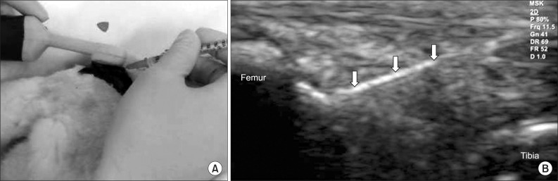
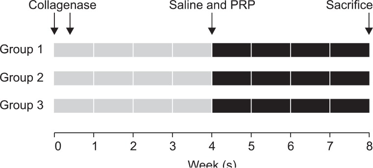
Anesthesia was induced with isoflurane (JW Pharmaceutical, Goyang, Korea) vaporized in oxygen and delivered via a large animal circle system. Both knee joints of all rabbits were shaved and sterilized, and three different doses (0.25 mg, 0.5 mg, and 1.0 mg) of collagenase type II from Clostridium histolyticum (Sigma-Aldrich, St. Louis, USA) were prepared for an intra-articular knee injection. Collagenase was dissolved in a sterile phosphate buffered saline (pH 7.4), and was filtered through a 0.22 µm membrane. The 21 rabbits were equally divided into three groups of seven. Injections of 0.25 mg, 0.5 mg, and 1.0 mg collagenase were delivered to both knees of group 1, 2, and 3, respectively. All injections were conducted by a physiatrist, using a commercially available ultrasound system E-CUBE 9® 3-12 MHz multi-frequency linear transducer (Alpionion Medical Systems, Seoul, Korea) (Fig. 1). The second injection with the same collagenase dose was repeated 3 days later (Fig. 2).10,11
PRP preparation, platelet count analysis, and regimen administration
For PRP preparation, 6.0 ml venous blood was drawn, using an aseptic technique from the marginal auricular vein and mixed with 2 ml of 0.129-mol/L sodium citrate in a sPRP system® PRP device (Huons, Goyang, Korea). It was centrifuged for 3 minutes at 3,200 rpm. The three layers that formed were comprised of platelet poor plasma, PRP, and red blood cells. The PRP layer was extracted through a special port and 0.3 ml was used for the injections. The number of platelets in the whole blood and isolated PRP fraction was assessed, using the Vet abc Plus hematology analyzer (Scil Animal Care, IL, USA). The mean platelet concentration of the whole blood was 324±25×103/µl (range: 277-356×103/µl). The mean platelet concentration of the PRP fraction was 2,664±970×103/µl (range: 2,005-3,120×103/µl), and represented an 8.2-fold increase over the concentration in the whole blood.
PRP (0.3 ml) was injected into the left knee and saline (0.3 ml) into the right knee, in rabbits in the three groups under ultrasound guidance, 4 weeks after the first collagenase injection.11,12 All procedures were performed under general anesthesia and sterile conditions. No medication was administered after the injection. The rabbits were euthanized by CO inhalation at 9 weeks after the first collagenase injection (Fig. 2).
Lameness of the affected limb
Clinical observations were performed once daily in the afternoon. The rabbits were turned out on the ground of the area of 2 square meters, and their gait pattern was assessed by direct observation for 20 min, individually. In the intact limb, the knee and ankle angles went through a typical flexion and extension cycle during hoping of the rabbit. The lameness was defined as non-weight bearing of the affected limb and losing of typical flexion and extension cycle during hoping in comparison with the unaffected limb. The severity of the lameness was not quantified. The times to normal ambulation without the non-weight bearing lameness of the affected limb were recorded, and the lameness periods were calculated for each group. Two independent physiatrists, without prior knowledge of the experimental groups, performed the observation.
Gross morphologic observations and macroscopic score
The knee joints were dissected after euthanasia and the lateral femoral condyle, which was the most destructive portion by collagenase in the knee, was examined for gross morphologic changes.10 Cartilage degeneration was assessed and measured using the Yoshimi scoring system (0, normal cartilage; 1, softened cartilage; 2, fibrillation; 3, erosion; 4, ulceration; and 5, loss of cartilage).13
Histological examination and score
The lateral femoral condyle was fixed with 10% neutral buffered formalin and decalcified with 20% ethylenediaminetetraacetic acid. The calcified condyle was embedded in paraffin and the standard frontal microsections with 5 µm width were prepared and stained with hematoxylin and eosin. If the stain was not proper, the next cartilage surface of the specimen was cut and stained. The degree of the cartilage degradation was assessed using the scoring system modified by Mankin et al.14 Histological evidence of cartilage degeneration was evaluated by the structural change of articular cartilage (0, normal; 1, surface irregularities; 2, pannus and surface irregularities; 3, clefts to transitional zones; 4, clefts to radial zones; 5, clefts to calcified zones; and 6, complete disorganization) and the cell status (0, normal; 1, diffuse hypercellularity; 2, cloning; and 3, hypocellularity). Total cartilage degeneration score ranged from 0 (normal) to 9 (complete disorganization and hypocellularity of articular cartilage). All cartilage sections were graded by a pathologist who was kept unaware of the treatment.
Statistical analyses
Statistical analyses were performed using the SPSS V. 14.0 software (SPSS, Chicago, USA). Kruskal-Wallis test was used to compare the gross and histological changes of cartilage among three groups with saline injection, and the difference of gross morphologic and histologic scores between saline- and PRP-injected knees among the three groups. Wilcoxon signed rank test was used to assess the differences between both knees injected with PRP and saline in each group. p-value<0.05 was considered statistically significant.
Lameness period of the affected limb
The average time to recovery of normal ambulation was 14±2 days in group 1 rabbits, 16±3 days in group 2 rabbits, and 20±3 days in group 3 rabbits. The average time to recovery of normal ambulation of group 3 was significantly longer than that of group 1 and 2 (p<0.05).
Gross morphologic and histologic findings according to the injection dose of collagenase in saline injected knees
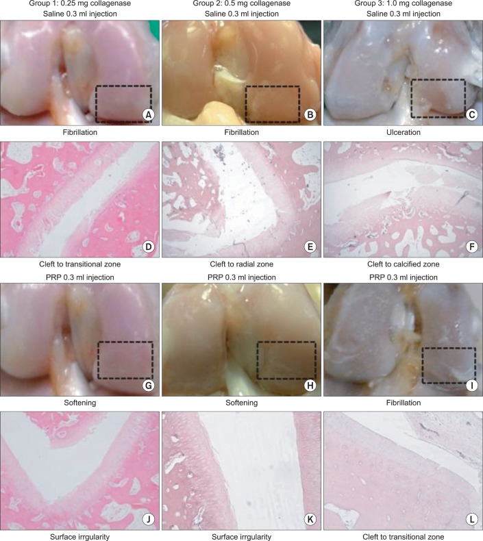
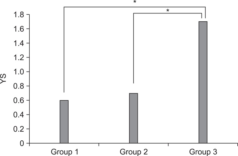
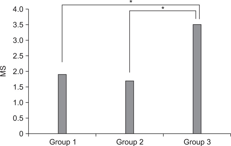
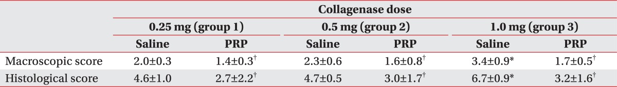
The severity of cartilage degeneration at the lateral femoral condyle was proportional to the increase of the injection dose of collagenase. Macroscopic and histological scores in group 3 were significantly higher than in groups 1 and 2 (p<0.05) (Table 1). However, there was no difference of scores between groups 1 and 2. The abnormal gross morphologic and histologic changes, such as the fibrillation and irregularity of cartilage surfaces, disappearance of surface-layer cells, and slightly diffuse cell growth in the transitional and radial zones were observed in group 1 (Fig. 3-A, D). Fibrillation of articular cartilage, and the cleft and cell cloning in the transitional and radial zones were noted in group 2 (Fig. 3-B, E). The ulceration of cartilage surface and loss and cell cloning in the radial zone were evident in group 3 (Fig. 3-C, F).
Comparison of gross morphologic and histologic findings between saline- and PRP-injected knees
Macroscopic and histological scores of the PRP-injected knees were significantly lower than those of the saline-injected knees in all groups (p<0.05) (Table 1). The difference of gross morphologic and histologic scores between the saline and PRP-injected knees in group 3 was higher than those in groups 1 and 2 (p<0.05) (Fig. 4, 5). However, there was no significant difference between groups 1 and 2. Histologic examination showed nearly normal cartilage surfaces with normal cellularity in the transitional and radial zones in group 1 with PRP injection, irregularity of cartilage surface with normal cellularity in the transitional zone in group 2 with PRP injection, and a cleft in the transitional with apparent cloning of chondrocytes in the transitional zone in group 3 with PRP injection (Fig. 3-J, L).
Our study reveals that the difference of gross morphologic and histologic scores between the saline and PRP injected knees in group 3 was higher than those in groups 1 and 2, which suggests that PRP injection is more regenerative in the 1.0 mg collagenase-induced knee OA, like group 3, than the other groups. It is postulated that a comparatively lesser cartilage regeneration happened in groups 1 and 2 than in group 3, because the degree of cartilage degeneration in groups 1 and 2 was less than that in group 3, after an intra-articular collagenase injection. These findings are consistent with a previous study that the OA changes were induced by the intra-articular injection of different doses of collagenase, and were dependent on the injection doses.13 Additionally, the potent cartilage protective effects of several therapeutic regimens were proved in a 1.0 mg collagenase-induced osteoarthritis rabbit model. Therefore, we speculate that 1.0 mg collagenase injection is sufficient to induce reversible OA-like changes.
Some recent animal studies assessed the PRP effect on chondrogenesis and cartilage healing. In a rabbit model, 48 osteochondral defects, created in the femoropatellar groove, were divided into three groups: no treatment, treatment with autogenous PRP in poly-lacticglycolic acid, and with poly-lacticglycolic acid alone.8 The PRP treatment group demonstrated a greater extent of cartilage regeneration, as well as higher production of glycosaminoglycans in the extracelluar matrix. In addition, chondrogenesis was also verified in cartilage defects of the rabbit knee when PRP was used with a scaffold.15 Increased hyaluronic acid production and secretion were detected in synoviocytes from the patients with OA cultured in PRP. This suggests that intra-articular PRP injection could potentially serve as the endogenous source of chondroprotection and joint lubrication.16 Our results are similar to those of a previous study that reported PRP-mediated stimulation of either cell proliferation or matrix metabolism through articular chondrocytes in vitro.17 To the best of our knowledge, this is the first study to assess the possibility that the severity of knee OA may have an impact on the therapeutic response of intra-articular PRP injection.
Animal models of OA, in which an articular cartilage damage is induced with intra-articular collagenase injection, have proven to be similar to human OA.13 Presently, in a rabbit model of OA of the knee, the degree of articular cartilage degeneration at the lateral femoral condyle was proportional to the increase of intra-articular injection dose of collagenase. These findings are consistent with a previous study that the OA changes were induced by the intra-articular injection of 0.5-2.0 mg collagenase, and were dependent on the injection doses.13 In addition, presently, knee OA injected with PRP showed significantly lower gross and histological scores than those injected with saline, indicating that PRP injection facilitates the regeneration of damaged cartilage in collagenase-induced knee OA. There are several explanations for the healing effect of PRP injection in knee OA. First, several growth factors are usually stored in alpha-granules of platelets as the latent form. When platelets are activated, these factors are gradually released and participate in the regeneration process.18 Second, chondrocytes derived from the surrounding normal cartilage may be activated, and then migrate into the damaged cartilage. Third, mesenchymal stem cells or other multi-potent stem cells may also become activated and migrate to the cartilage lesion. This process is corroborated by a previous study that demonstrated the chondrogenic and proliferative effects of PRP on mesenchymal stem cells.19
The ideal concentration of platelets in PRP is not yet clear. Previous study demonstrated that the concentrations of growth factors in PRP were positively correlated with that of the platelet concentration of PRP.20 Qualitative and quantitative changes of the platelets may affect the regenerative power of PRP. In our study, PRP was prepared from the centrifugation of the autologous whole blood in rabbits, and the platelet concentration of the whole blood was compatible with the range of normal values for rabbit (158-650×103/µl).21 The platelet concentration of PRP was 8.2-times higher than that of the whole blood, thus, offering higher regenerative effects of PRP. Therefore, our PRP preparation is adequate to assess its therapeutic effects for cartilage regeneration because of the high platelet concentration.
Until now, intra-articular injection into the rabbit knee, under ultrasound guidance, has not been reported. In a human study, one third of the blind knee injections, performed by physician, were shown to be inaccurate.22 In our study, improvement of accuracy in the knee injection was achieved through an ultrasound guidance. A transducer was placed transversally on the anteriolateral portion of the rabbit knee, and intra-articular injection was performed when a needle was correctly placed in the knee joint on ultrasound guidance.
There are several limitations in our study. First, the small number of specimens made it difficult to determine the therapeutic effect of PRP in the knee OA of rabbits. Second, there was no control group for the comparison of the cartilage degeneration effects, according to the injection dose of collagenase, because specimens were not examined at 4 weeks after the first collagenase injection. Third, we did not conduct the histological examination of synovial changes in our study. The synovial membrane is also a source of proinflammatory and catabolic products, including metalloproteinases and aggrecanases, which contribute to articular matrix degradation. In the previous study, the inflammatory synovial reaction of the knee joint was the most severe at 1 week after collagenase injection. After then, the inflammatory responses in the synovial tissue lessened with time and weekly remained at 6 weeks after injection.13 In our study, PRP was injected into the knee at 4 weeks after the first collagenase injection, and the rabbits were euthanized at 9 weeks after the first injection. Therefore, it was difficult to decide whether the synovial changes were due to the effect of PRP or simply as the passing of time. Fourth, we did not consider the influence of physical performance of rabbit on cartilage regeneration. Fifth, we did not contemplate the influence experimental rabbit age since the degenerative processes progress, as time passed and better results are achieved in younger rabbits. Finally, we did not evaluate the therapeutic effects of PRP in the severe knee OA. Further studies will be needed to evaluate the PRP effects, according to the different platelet concentration, injection time, and number of injection in knee OA of rabbits, and in severe knee OA for the achievement of the best and more durable results.
Although our results are based on a relatively small number of specimens, the degree of cartilage degeneration was directly proportional to the collagenase injection dose in the knee OA of rabbits, and PRP injection demonstrated the effect of cartilage regeneration in all severity of rabbits' knee OA. In addition, the cartilage regenerative power of PRP injection in medium dose collagenase induced knee OA was greater than that in low doses. Further formal preclinical trial with more specimens is required to establish the validity of our study.
  • 1. Wearing SC, Hennig EM, Byrne NM, Steele JR, Hills AP. Musculoskeletal disorders associated with obesity: a biomechanical perspective. Obes Rev 2006;7:239-250.
  • 2. Hochberg MC, Altman RD, Brandt KD, Clark BM, Dieppe PA, Griffin MR, Moskowitz RW, Schnitzer TJ. Guidelines for the medical management of osteoarthritis. Part II. Osteoarthritis of the knee. American College of Rheumatology. Arthritis Rheum 1995;38:1541-1154.
  • 3. Goldspink DF, Goldberg AL. Influence of pituitary growth hormone on DNA synthesis in rat tissues. Am J Physiol 1975;228:302-309.
  • 4. Altman R, Brandt K, Hochberg M, Moskowitz R, Bellamy N, Bloch DA, Buckwalter J, Dougados M, Ehrlich G, Lequesne M, et al. Design and conduct of clinical trials in patients with osteoarthritis: recommendations from a task force of the Osteoarthritis Research Society. Results from a workshop. Osteoarthritis Cartilage 1996;4:217-243.
  • 5. Namiki O, Toyoshima H, Morisaki N. Therapeutic effect of intra-articular injection of high molecular weight hyaluronic acid on osteoarthritis of the knee. Int J Clin Pharmacol Ther Toxicol 1982;20:501-507.
  • 6. Anitua E, Sanchez M, Nurden AT, Nurden P, Orive G, Andia I. New insights into and novel applications for platelet-rich fibrin therapies. Trends Biotechnol 2006;24:227-234.
  • 7. Sampson S, Gerhardt M, Mandelbaum B. Platelet rich plasma injection grafts for musculoskeletal injuries: a review. Curr Rev Musculoskelet Med 2008;1:165-174.
  • 8. Sun Y, Feng Y, Zhang CQ, Chen SB, Cheng XG. The regenerative effect of platelet-rich plasma on healing in large osteochondral defects. Int Orthop 2010;34:589-597.
  • 9. Schmidt MB, Chen EH, Lynch SE. A review of the effects of insulin-like growth factor and platelet derived growth factor on in vivo cartilage healing and repair. Osteoarthritis Cartilage 2006;14:403-412.
  • 10. Kikuchi T, Sakuta T, Yamaguchi T. Intra-articular injection of collagenase induces experimental osteoarthritis in mature rabbits. Osteoarthritis Cartilage 1998;6:177-186.
  • 11. Kim SB, Kwon DR, Kwak H, Shin YB, Han HJ, Lee JH, Choi SH. Additive effects of intra-articular injection of growth hormone and hyaluronic acid in rabbit model of collagenase-induced osteoarthritis. J Korean Med Sci 2010;25:776-780.
  • 12. Huh JE, Baek YH, Lee JD, Choi DY, Park DS. Therapeutic effect of Siegesbeckia pubescens on cartilage protection in a rabbit collagenase-induced model of osteoarthritis. J Pharmacol Sci 2008;107:317-328.
  • 13. Yoshimi T, Kikuchi T, Obara T, Yamaguchi T, Sakakibara Y, Itoh H, Iwata H, Miura T. Effects of high-molecular-weight sodium hyaluronate on experimental osteoarthrosis induced by the resection of rabbit anterior cruciate ligament. Clin Orthop Relat Res 1994;298:296-304.
  • 14. Mankin HJ, Dorfman H, Lippiello L, Zarins A. Biochemical and metabolic abnormalities in articular cartilage from osteo-arthritic human hips. II. Correlation of morphology with biochemical and metabolic data. J Bone Joint Surg Am 1971;53:523-537.
  • 15. Qi YY, Chen X, Jiang YZ, Cai HX, Wang LL, Song XH, Zou XH, Ouyang HW. Local delivery of autologous platelet in collagen matrix simulated in situ articular cartilage repair. Cell Transplant 2009;18:1161-1169.
  • 16. Anitua E, Sanchez M, Nurden AT, Zalduendo MM, de la Fuente M, Azofra J, Andia I. Platelet-released growth factors enhance the secretion of hyaluronic acid and induce hepatocyte growth factor production by synovial fibroblasts from arthritic patients. Rheumatology 2007;46:1769-1772.
  • 17. Akeda K, An HS, Okuma M, Attawia M, Miyamoto K, Thonar EJ, Lenz ME, Sah RL, Masuda K. Platelet-rich plasma stimulates porcine articular chondrocyte proliferation and matrix biosynthesis. Osteoarthritis Cartilage 2006;14:1272-1280.
  • 18. Marx RE. Platelet-rich plasma: evidence to support its use. J Oral Maxillofac Surg 2004;62:489-496.
  • 19. Drengk A, Zapf A, Sturmer EK, Sturmer KM, Frosch KH. Influence of platelet-rich plasma on chondrogenic differentiation and proliferation of chondrocytes and mesenchymal stem cells. Cells Tissues Organs 2009;189:317-326.
  • 20. Li M, Zhang C, Yuan T, Chen S, Lu R. Assessment study on a set of platelet-rich plasma preparation. Zhongguo Xiu Fu Chong Jian Wai Ke Za Zhi 2011;25:112-116.
  • 21. Fox JG, Anderson LC, Loew FM, Quimby FW. Laboratory animal medicine. 2002.2nd ed. Salt Lake: Academic Press.
  • 22. Cunnington J, Marshall N, Hide G, Bracewell C, Isaacs J, Platt P, Kane D. A randomized, double-blind, controlled study of ultrasound-guided corticosteroid injection into the joint of patients with inflammatory arthritis. Arthritis Rheum 2010;62:1862-1869.
Fig. 1
(A) Intra-articular injection was performed into a knee of rabbit under ultrasound guidance. (B) Longitudinal ultrasound image showed the needle (arrows) in the knee joint of rabbit.
arm-36-458-g001.jpg
Fig. 2
Time line of collagenase, saline, and PRP injection. Both knees injections were performed twice with 0.25 mg (group 1), 0.5 mg (group 2), and 1.0 mg (group 3) collagenase on day 1 and 4, respectively. 0.3 ml PRP was injected into the left knee and 0.3 ml saline into the right knee in three groups at 4 weeks after the first collagenase injection. All rabbits were euthanized by CO inhalation at 4 weeks after saline and PRP injections.
arm-36-458-g002.jpg
Fig. 3
Gross morphological (rectangular areas) and histological (hematoxylin and eosin staining, ×40) findings of knee osteoarthritis in group 1, 2, and 3 with intra-articular injection of saline (A-F) and platelet rich plasma (G-L).
arm-36-458-g003.jpg
Fig. 4
Comparison of differences in gross morphological score between saline and PRP injected knees among three groups. YS: Yoshimi's score. *p<0.05 by Kruskall-Wallis test.
arm-36-458-g004.jpg
Fig. 5
Comparison of differences in histological evaluation score between saline and PRP Injected knees among three groups. MS: modified Mankin's score. *p<0.05 by Kruskall-Wallis test.
arm-36-458-g005.jpg
Table 1
Macroscopic and Histological Scores in Collagenase-Induced Knee Osteoarthritis of Rabbits with Intra-Articular injection of Saline and Platelet-Rich Plasma

Values are mean±standard deviation

PRP: Platelet-rich plasma

*p<0.05 among 3 groups with saline injection, p<0.05 between saline and PRP

arm-36-458-i001.jpg

Figure & Data

References

    Citations

    Citations to this article as recorded by  
    • Evaluating the efficacy of intra-articular polydioxanone (PDO) injections as a novel viscosupplement in osteoarthritis treatment
      Linh Thi Thuy Le, Pham Ngoc Chien, Thuy-Tien Thi Trinh, Ji-Won Seo, Nguyen Ngan Giang, Pham Thi Nga, Xin Rui Zhang, Yong Xun Jin, Sun-Young Nam, Chan-Yeong Heo
      Life Sciences.2025; 361: 123303.     CrossRef
    • Chondroprotective Effects of a Single PRP Injection in a Spontaneous Osteoarthritis Model of Dunkin Hartley Guinea Pig: An Immunohistochemical Analysis
      Devendra Kumar Chouhan, Sandeep Patel, Tarkik Thami, Narayan Prasad Mishra, Uma Nahar, Mandeep Singh Dhillon
      Indian Journal of Orthopaedics.2024; 58(7): 887.     CrossRef
    • Mechanisms and applications of the regenerative capacity of platelets-based therapy in knee osteoarthritis
      Jiang-Yin Zhang, Xiao-Na Xiang, Xi Yu, Yan Liu, Hong-Ying Jiang, Jia-Lei Peng, Cheng-Qi He, Hong-Chen He
      Biomedicine & Pharmacotherapy.2024; 178: 117226.     CrossRef
    • Donor-Matched Peripheral Blood–Derived Mesenchymal Stem Cells Combined With Platelet-Rich Plasma Synergistically Ameliorate Surgery-Induced Osteoarthritis in Rabbits: An In Vitro and In Vivo Study
      Kaibo Zhang, Tianhao Xu, Huiqi Xie, Jian Li, Weili Fu
      The American Journal of Sports Medicine.2023; 51(11): 3008.     CrossRef
    • Liquid platelet-rich fibrin produced via horizontal centrifugation decreases the inflammatory response and promotes chondrocyte regeneration in vitro
      Huimin Li, Ting Xia, Hao Zeng, Yun Qiu, Yan Wei, Yihong Cheng, Yulan Wang, Xiaoxin Zhang, Jin Ke, Richard Miron, Qing He
      Frontiers in Bioengineering and Biotechnology.2023;[Epub]     CrossRef
    • Role of platelet rich plasma in management of early knee osteoarthritis pain: A retrospective observational study
      Sandeep Khuba, Dheeraj Khetan, Sanjay Kumar, Keshav Kumar Garg, Sujeet Gautam, Prabhaker Mishra
      Interventional Pain Medicine.2023; 2(4): 100297.     CrossRef
    • The efficacy of transcatheter arterial embolization for knee pain on patients with knee osteoarthritis: A case series
      Kun Yung Kim, Gi-Wook Kim
      Journal of Back and Musculoskeletal Rehabilitation.2022; 35(4): 743.     CrossRef
    • Chondroprotective effects of multiple PRP injections in osteoarthritis by apoptosis regulation and increased aggrecan synthesis- Immunohistochemistry based Guinea pig study
      Sandeep Patel, Narayan Prasad Mishra, Devendra Kumar Chouhan, Uma Nahar, Mandeep S. Dhillon
      Journal of Clinical Orthopaedics and Trauma.2022; 25: 101762.     CrossRef
    • Clinical benefit of high tibial osteotomy combined with the intervention of platelet-rich plasma for severe knee osteoarthritis
      Conglei Dong, Chao Zhao, Fei Wang
      Journal of Orthopaedic Surgery and Research.2022;[Epub]     CrossRef
    • A novel cell-free intrathecal approach with PRP for the treatment of spinal cord multiple sclerosis in cats
      Mariam F. Farid, Yara S. Abouelela, Noha A. E. Yasin, Mohamed R. Mousa, Marwa A. Ibrahim, Abdelbary Prince, Hamdy Rizk
      Inflammation and Regeneration.2022;[Epub]     CrossRef
    • A Combination of Surgical and Chemical Induction in a Rabbit Model for Osteoarthritis of the Knee
      Eun Jeong Go, Seon Ae Kim, Mi-La Cho, Kwan Soo Lee, Asode Ananthram Shetty, Seok Jung Kim
      Tissue Engineering and Regenerative Medicine.2022; 19(6): 1377.     CrossRef
    • PRP Injections for the Treatment of Knee Osteoarthritis: A Meta-Analysis of Randomized Controlled Trials
      Giuseppe Filardo, Davide Previtali, Francesca Napoli, Christian Candrian, Stefano Zaffagnini, Alberto Grassi
      CARTILAGE.2021; 13(1_suppl): 364S.     CrossRef
    • Potential Mechanism of Action of Current Point-of-Care Autologous Therapy Treatments for Osteoarthritis of the Knee—A Narrative Review
      Jennifer Woodell-May, Kathleen Steckbeck, William King
      International Journal of Molecular Sciences.2021; 22(5): 2726.     CrossRef
    • Impact of platelet-rich plasma versus selenium in ameliorating induced toxicity in rat testis: histological, immunohistochemical, and molecular study
      Walaa Mohamed Sayed, Ahmed Elzainy
      Cell and Tissue Research.2021; 385(1): 223.     CrossRef
    • T2-mapping MRI evaluation of patellofemoral cartilage in patients submitted to intra-articular platelet-rich plasma (PRP) injections
      Flavia Cobianchi Bellisari, Luigi De Marino, Francesco Arrigoni, Silvia Mariani, Federico Bruno, Pierpaolo Palumbo, Camilla De Cataldo, Ferruccio Sgalambro, Nadia Catallo, Luigi Zugaro, Ernesto Di Cesare, Alessandra Splendiani, Carlo Masciocchi, Andrea Gi
      La radiologia medica.2021; 126(8): 1085.     CrossRef
    • Platelet-rich plasma injections induce disease-modifying effects in the treatment of osteoarthritis in animal models
      Angelo Boffa, Manuela Salerno, Giulia Merli, Laura De Girolamo, Lior Laver, Jérémy Magalon, Mikel Sánchez, Thomas Tischer, Giuseppe Filardo
      Knee Surgery, Sports Traumatology, Arthroscopy.2021; 29(12): 4100.     CrossRef
    • Effects of platelet-rich plasma on the clinical outcomes and cartilage thickness in patients with knee osteoarthritis
      Ekin İlke Şen, Mustafa Aziz Yıldırım, Tuğba Yeşilyurt, Fatma Nur Kesiktaş, Demirhan Dıraçoğlu
      Journal of Back and Musculoskeletal Rehabilitation.2020; 33(4): 597.     CrossRef
    • Orthobiologics for the Hip Region: A Narrative Review
      Kelly C. McInnis, Eric T. Chen, Jonathan T. Finnoff, Eugene Y. Roh, Joanne Borg Stein
      PM&R.2020; 12(10): 1045.     CrossRef
    • Local Injection of Growth Hormone for Temporomandibular Joint Osteoarthritis
      Soo-Min Ok, Jin-Hwa Kim, Ji-Su Kim, Eun-gyo Jeong, Yang Mi Park, Hye-Mi Jeon, Jun-Young Heo, Yong-Woo Ahn, Sun-Nyoung Yu, Hae Ryoun Park, Kyung-Hee Kim, Soon-Cheol Ahn, Sung-Hee Jeong
      Yonsei Medical Journal.2020; 61(4): 331.     CrossRef
    • Neuroimmune modulation of pain and regenerative pain medicine
      Thomas Buchheit, Yul Huh, William Maixner, Jianguo Cheng, Ru-Rong Ji
      Journal of Clinical Investigation.2020; 130(5): 2164.     CrossRef
    • Nanomechanical Properties of Articular Cartilage Due to the PRP Injection in Experimental Osteoarthritis in Rabbits
      Mikhail Ihnatouski, Jolanta Pauk, Boris Karev, Dmitrij Karev
      Molecules.2020; 25(16): 3734.     CrossRef
    • Ortho-Biologics for Osteoarthritis
      Kyla Huebner, Rachel M. Frank, Alan Getgood
      Clinics in Sports Medicine.2019; 38(1): 123.     CrossRef
    • Intra-articular Recombinant Human Growth Hormone Injection Compared with Hyaluronic Acid and Placebo for an Osteoarthritis Model of New Zealand Rabbits
      Andri Maruli Tua Lubis, Erick Wonggokusuma, Aldo Fransiskus Marsetio
      Knee Surgery and Related Research.2019; 31(1): 44.     CrossRef
    • In Vitro Evaluation of Small Molecule Delivery into Articular Cartilage: Effect of Synovial Clearance and Compressive Load
      Tim W.G.M. Spitters, Dimitrios Stamatialis, Audrey Petit, Mike G.W. de Leeuw, Marcel Karperien
      ASSAY and Drug Development Technologies.2019; 17(4): 191.     CrossRef
    • Multiple Platelet-Rich Plasma Injections Versus Single Platelet-Rich Plasma Injection in Early Osteoarthritis of the Knee: An Experimental Study in a Guinea Pig Model of Early Knee Osteoarthritis
      Devendra K. Chouhan, Mandeep S. Dhillon, Sandeep Patel, Tungish Bansal, Alka Bhatia, Himanshu Kanwat
      The American Journal of Sports Medicine.2019; 47(10): 2300.     CrossRef
    • Is local platelet-rich plasma injection clinically superior to hyaluronic acid for treatment of knee osteoarthritis? A systematic review of randomized controlled trials
      Yalong Di, Changxu Han, Liang Zhao, Yizhong Ren
      Arthritis Research & Therapy.2018;[Epub]     CrossRef
    • Role of autophagy in the progression of osteoarthritis: The autophagy inhibitor, 3-methyladenine, aggravates the severity of experimental osteoarthritis
      Ni-Tao Cheng, Hai Meng, Li-Feng Ma, Liang Zhang, Hao-Miao Yu, Zhen-Zhong Wang, Ai Guo
      International Journal of Molecular Medicine.2017; 39(5): 1224.     CrossRef
    • Platelet-Rich Plasma for the Management of Hip and Knee Osteoarthritis
      Kim L. Bennell, David J. Hunter, Kade L. Paterson
      Current Rheumatology Reports.2017;[Epub]     CrossRef
    • Reconstruction of cranial cruciate ligament in rabbits using polyester implants saturated with PRP, antlerogenic stem cells MIC-1 and their homogenate
      Przemysław Prządka, Zdzisław Kiełbowicz, Bogdan Osiński, Stanisław Dzimira, Janusz A. Madej, Wojciech Nowacki, Krzysztof Kubiak, Paweł Reichert, Marek Cegielski
      Connective Tissue Research.2017; 58(5): 464.     CrossRef
    • Nonsurgical Treatment of Ankle Arthritis
      Michael A. Gentile
      Clinics in Podiatric Medicine and Surgery.2017; 34(4): 415.     CrossRef
    • Role of PRP in the Treatment of Knee Osteoarthritis
      John S. Georgy, Lawrence P. Lai, Todd P. Stitik, Raj D. Desai, Charles Koon, Sanjay Kumbar, Boqing Chen, Patrick M. Foye
      Current Physical Medicine and Rehabilitation Reports.2016; 4(2): 138.     CrossRef
    • Does platelet-rich plasma have a role in the treatment of osteoarthritis?
      Paul Ornetti, Geoffroy Nourissat, Francis Berenbaum, Jérémie Sellam, Pascal Richette, Xavier Chevalier
      Joint Bone Spine.2016; 83(1): 31.     CrossRef
    • Evidence and recommendations for use of intra-articular injections for knee osteoarthritis
      Christelle Nguyen, Marie-Martine Lefèvre-Colau, Serge Poiraudeau, François Rannou
      Annals of Physical and Rehabilitation Medicine.2016; 59(3): 184.     CrossRef
    • MORPHOGENESIS OF KNEE HYALINE CARTILAGE DURING INTRAARTICULAR INJECTION OF PLATELET-RICH AUTOLOGOUS PLASMA AND/OR HYALURONIC ACID PREPARATION IN RATS WITH EXPERIMENTAL OSTEOARTHRITIS
      S. A. Demkin, D. A. Malanin, L. N. Rogova, G. L. Snigur, N. V. Grigorieva, K. V. Baydova
      Traumatology and Orthopedics of Russia.2016; 22(4): 76.     CrossRef
    • Biologic Strategies for Intra-articular Treatment and Cartilage Repair
      Susana O. Monteiro, Elisa V. Bettencourt, Olivier M. Lepage
      Journal of Equine Veterinary Science.2015; 35(3): 175.     CrossRef
    • Platelet-rich plasma: why intra-articular? A systematic review of preclinical studies and clinical evidence on PRP for joint degeneration
      G. Filardo, E. Kon, A. Roffi, B. Di Matteo, M. L. Merli, M. Marcacci
      Knee Surgery, Sports Traumatology, Arthroscopy.2015; 23(9): 2459.     CrossRef
    • Platelet-rich plasma to treat ankle cartilage pathology - from translational potential to clinical evidence: a systematic review
      Francesca Vannini, Berardo Di Matteo, Giuseppe Filardo
      Journal of Experimental Orthopaedics.2015;[Epub]     CrossRef
    • Differentiation Effects of Platelet-Rich Plasma Concentrations on Synovial Fluid Mesenchymal Stem Cells from Pigs Cultivated in Alginate Complex Hydrogel
      Hao-Che Tang, Wei-Chuan Chen, Chih-Wei Chiang, Lei-Yen Chen, Yu-Ching Chang, Chih-Hwa Chen
      International Journal of Molecular Sciences.2015; 16(8): 18507.     CrossRef
    • Intraarticular injections (corticosteroid, hyaluronic acid, platelet rich plasma) for the knee osteoarthritis
      Egemen Ayhan
      World Journal of Orthopedics.2014; 5(3): 351.     CrossRef
    • Knee osteoarthritis: hyaluronic acid, platelet-rich plasma or both in association?
      Isabel Andia, Michele Abate
      Expert Opinion on Biological Therapy.2014; 14(5): 635.     CrossRef
    • Biology of platelet-rich plasma and its clinical application in cartilage repair
      Xuetao Xie, Changqing Zhang, Rocky S Tuan
      Arthritis Research & Therapy.2014;[Epub]     CrossRef
    • The Use of Platelet-Rich Plasma for the Treatment of Osteoarthritis
      Prakash Jayabalan, Sarah Hagerty, Megan Helen Cortazzo
      The Physician and Sportsmedicine.2014; 42(3): 53.     CrossRef
    • Expression of ADAMTs-5 and TIMP-3 in the condylar cartilage of rats induced by experimentally created osteoarthritis
      Wen Li, Mengjie Wu, Shijie Jiang, Wanghui Ding, Qiaojie Luo, Jiejun Shi
      Archives of Oral Biology.2014; 59(5): 524.     CrossRef
    • Platelet-rich plasma limits the nerve injury caused by 10% dextrose in the rabbit median nerve
      Gi-Young Park, Dong Rak Kwon
      Muscle & Nerve.2014; 49(1): 56.     CrossRef
    • Drug delivery systems for intra-articular treatment of osteoarthritis
      Mi Lan Kang, Gun-Il Im
      Expert Opinion on Drug Delivery.2014; 11(2): 269.     CrossRef
    • Quelle place pour les PRP (plasma riche en plaquettes) dans l’arthrose ?
      Paul Ornetti, Geoffroy Nourissat, Francis Berenbaum, Jérémie Sellam, Pascal Richette, Xavier Chevalier
      Revue du Rhumatisme.2014; 81(6): 466.     CrossRef
    • The Use of Platelet-Rich Plasma Preparations in the Treatment of Musculoskeletal Injuries in Orthopaedic Sports Medicine
      Simone Cerciello, Knut Beitzel, Nathan Howlett, Ryan P. Russell, John Apostolakos, Mary Beth McCarthy, Mark P. Cote, Augustus D. Mazzocca
      Operative Techniques in Orthopaedics.2013; 23(2): 69.     CrossRef
    • Basic science and clinical application of platelet-rich plasma for cartilage defects and osteoarthritis: a review
      Y. Zhu, M. Yuan, H.Y. Meng, A.Y. Wang, Q.Y. Guo, Y. Wang, J. Peng
      Osteoarthritis and Cartilage.2013; 21(11): 1627.     CrossRef
    • Activated platelet-rich plasma improves adipose-derived stem cell transplantation efficiency in injured articular cartilage
      Phuc Van Pham, Khanh Hong-Thien Bui, Dat Quoc Ngo, Ngoc Bich Vu, Nhung Hai Truong, Nhan Lu-Chinh Phan, Dung Minh Le, Triet Dinh Duong, Thanh Duc Nguyen, Vien Tuong Le, Ngoc Kim Phan
      Stem Cell Research & Therapy.2013;[Epub]     CrossRef

    Download Citation

    Download a citation file in RIS format that can be imported by all major citation management software, including EndNote, ProCite, RefWorks, and Reference Manager.

    Format:

    Include:

    The Effects of Intra-Articular Platelet-Rich Plasma Injection According to the Severity of Collagenase-Induced Knee Osteoarthritis in a Rabbit Model
    Ann Rehabil Med. 2012;36(4):458-465.   Published online August 27, 2012
    Download Citation
    Download a citation file in RIS format that can be imported by all major citation management software, including EndNote, ProCite, RefWorks, and Reference Manager.

    Format:
    • RIS — For EndNote, ProCite, RefWorks, and most other reference management software
    • BibTeX — For JabRef, BibDesk, and other BibTeX-specific software
    Include:
    • Citation for the content below
    The Effects of Intra-Articular Platelet-Rich Plasma Injection According to the Severity of Collagenase-Induced Knee Osteoarthritis in a Rabbit Model
    Ann Rehabil Med. 2012;36(4):458-465.   Published online August 27, 2012
    Close

    Figure

    • 0
    • 1
    • 2
    • 3
    • 4
    The Effects of Intra-Articular Platelet-Rich Plasma Injection According to the Severity of Collagenase-Induced Knee Osteoarthritis in a Rabbit Model
    Image Image Image Image Image
    Fig. 1 (A) Intra-articular injection was performed into a knee of rabbit under ultrasound guidance. (B) Longitudinal ultrasound image showed the needle (arrows) in the knee joint of rabbit.
    Fig. 2 Time line of collagenase, saline, and PRP injection. Both knees injections were performed twice with 0.25 mg (group 1), 0.5 mg (group 2), and 1.0 mg (group 3) collagenase on day 1 and 4, respectively. 0.3 ml PRP was injected into the left knee and 0.3 ml saline into the right knee in three groups at 4 weeks after the first collagenase injection. All rabbits were euthanized by CO inhalation at 4 weeks after saline and PRP injections.
    Fig. 3 Gross morphological (rectangular areas) and histological (hematoxylin and eosin staining, ×40) findings of knee osteoarthritis in group 1, 2, and 3 with intra-articular injection of saline (A-F) and platelet rich plasma (G-L).
    Fig. 4 Comparison of differences in gross morphological score between saline and PRP injected knees among three groups. YS: Yoshimi's score. *p<0.05 by Kruskall-Wallis test.
    Fig. 5 Comparison of differences in histological evaluation score between saline and PRP Injected knees among three groups. MS: modified Mankin's score. *p<0.05 by Kruskall-Wallis test.
    The Effects of Intra-Articular Platelet-Rich Plasma Injection According to the Severity of Collagenase-Induced Knee Osteoarthritis in a Rabbit Model

    Macroscopic and Histological Scores in Collagenase-Induced Knee Osteoarthritis of Rabbits with Intra-Articular injection of Saline and Platelet-Rich Plasma

    Values are mean±standard deviation

    PRP: Platelet-rich plasma

    *p<0.05 among 3 groups with saline injection, p<0.05 between saline and PRP

    Table 1 Macroscopic and Histological Scores in Collagenase-Induced Knee Osteoarthritis of Rabbits with Intra-Articular injection of Saline and Platelet-Rich Plasma

    Values are mean±standard deviation

    PRP: Platelet-rich plasma

    *p<0.05 among 3 groups with saline injection, p<0.05 between saline and PRP

    TOP