• KARM
  • Contact us
  • E-Submission
ABOUT
ARTICLE TYPES
BROWSE ARTICLES
AUTHOR INFORMATION

Articles

Original Article

Reliability of Hip Migration Index in Children with Cerebral Palsy: The Classic and Modified Methods

Annals of Rehabilitation Medicine 2012;36(1):33-38.
Published online: February 29, 2012

Department and Research Institute of Rehabilitation Medicine, Yonsei University College of Medicine, Seoul 120-752, Korea.

Corresponding author: Eun Sook Park. Department and Research Institute of Rehabilitation Medicine, Yonsei University College of Medicine, 134, Sinchon-dong, Seodaemun-gu, Seoul 120-752, Korea. Tel: +82-2-2228-3712, Fax: +82-2-363-2795, pes1234@yuhs.ac
• Received: August 1, 2011   • Accepted: October 26, 2011

Copyright © 2012 by Korean Academy of Rehabilitation Medicine

This is an Open Access article distributed under the terms of the Creative Commons Attribution Non-Commercial License (http://creativecommons.org/licenses/by-nc/3.0) which permits unrestricted noncommercial use, distribution, and reproduction in any medium, provided the original work is properly cited.

  • 8,139 Views
  • 54 Download
  • 33 Crossref
  • 33 Scopus
prev next
  • Objective
    To determine reliability and clinical use of two methods of migration index (MI) in CP patients with or without hip dysplasia.
  • Method
    The materials included radiographs of 200 hips of children with cerebral palsy. Conventional anteroposterior radiographs of the pelvis were taken with the child in the supine position with standardized methods. Two rehabilitation doctors measured the migration index using two methods. In the classic method, the lateral margin of the acetabular roof was used as a landmark and in the modified method the lateral margin of the sourcil was used as a landmark. Each rater measured the migration index at three separate times with a time interval of at least one week. Intraclass correlation (ICC) was used to test the inter- and intra-rater reliability.
  • Results
    MI shows excellent intra-rater reliability in both the classic and modified methods, but the inter-rater reliability was higher in the classic method than in the modified method. When categorized according to the sourcil classification, inter-rater reliability was higher in the normal sourcil type and lower in the dysplastic sourcil types.
  • Conclusion
    Generally, the classic method showed higher reliability than the modified method, even though the reliability of the MI measurement was relatively high with both methods.
The incidence rate of hip displacement is the highest after equinus in musculoskeletal deformities in patients with cerebral palsy. If appropriate therapeutic intervention is not applied, hip displacement may develop into hip dislocation and cause postural and gait abnormalities, scoliosis, hip joint pains and problems of perineal hygiene.1,2 The subluxation or dislocation of the hip joint is one of the complications faced by children with cerebral palsy, in whom the incidence rate reaches 15% to 30% for all cerebral palsy patients. It is especially higher in patients with quadriplegic cerebral palsy.
Early therapeutic intervention is known to reduce the incidence rate of serious hip displacement in children with cerebral palsy.3 Thus, the early detection of hip displacement is critical to delay the progress of hip displacement. Various radiologic indexes have been used to detect hip displacement in children with cerebral palsy early. This is particularly true for the migration index,4 which is the most frequently used index to evaluate the risk of hip displacement and associated therapeutic effects.
In previous studies, intra-rater reliabilities were high, but inter-rater reliabilities showed different results.5-7
In most cases, the lateral edge of the acetabular roof is used as the datum point in the process where the migration index is measured. In the case of acetabular dysplasia, however, the lateral edge of the acetabular roof may be irregular in its coordinates, and therefore the measured values may be different among raters.
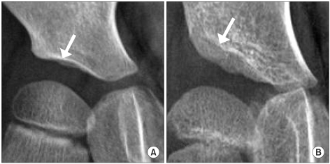
The sourcil is a shadow of dense osseous tissue that is observed at the weight-bearing surface of the acetabular roof (Fig. 1). In general, hip dysplasia can be evaluated by two methods, i.e., the classic method and the modified method. The former is to use the lateral edge of the acetabular roof as the datum point, and the latter is to use the lateral edge of the sourcil. In previous studies on the acetabular angles of patients who underwent operations for developmental hip displacement, it was reported that the modified method was more useful to evaluate hip dysplasia.8-10
Also in children with cerebral palsy, the measured value may be different according to datum points. Nevertheless, there has been no comparative study on the classic method and the modified method in relation to the migration index. This study analyzed intra-rater and inter-rater reliability not only in the classic method but in the modified method, as well as to study the clinical significance.
Subjects
This study was conducted on patients with cerebral palsy who visited the rehabilitation department at this hospital and underwent plain radiographies on their hip joints (200 hip joints) between August 2009 and December 2009. The study group consisted of 66 boys and 34 girls, whose average age was 7.88±1.54 years.
Methods
Subjects were assisted to take a supine position so their their pelvises could be parallel to each other and both their coxae could be in a neutral position while undergoing plain hip radiographies.11,12 This study was retrospectively conducted, and hip displacement was measured by two raters (A and B) using plain hip radiographs. The two raters were well-acquainted with pelvic and coxal structures and radiologic indexes, and measured hip displacement in accordance with standardized methods.4
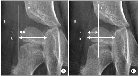
The outermost edge of the acetabular roof was regarded as the datum point from where the Perkin's line (P-line) was drawn. Likewise, another P-line was drawn from the lateral edge of the sourcil. The former and the latter method were defined as the classic and modified method, respectively (Fig. 2).
To evaluate intra-rater reliability, the displacement of 200 hip joints was measured 3 times at intervals of 1 week without data on previous measurements, and an analysis was made of the reliability amongst measured values. In order to evaluate inter-rater reliability, the displacement of 200 hip joints was measured in the same way, and an analysis was made of the reliability amongst mean values.
The reliabilities were evaluated in patients with or without hip dysplasia. In accordance with the descriptions of Ogata et al.13 and Agus et al.,8 the acetabular roof was classified as to the shape of the sourcil. Type I and II cases where the lateral edge of the acetabular roof and that of the sourcil are superimposed, were regarded as normal. On the other hand, type III and IV were classified as hip dysplasia.
Statistical analysis
Intra-rater reliability and inter-rater reliability were evaluated by use of Intraclass Correlation Coefficient (ICC), and the standard error of measurement (SEM) was calculated on the basis of inter-value differences and standard deviation. The data were analyzed by use of the program SPSS 18.0.
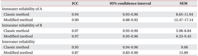
Intra-rater reliability was within reliable levels for both raters. In the case of rater-A, ICC indicated 0.94 and 0.90 in the classic method and the modified method, respectively. SEM indicated 8.64 to 11.94 and 12.48 to 17.14 respectively. In the case of rater-B, ICC was 0.97 in both methods. SEM indicated 5.98 to 8.84 and 6.23 to 9.45, respectively, and thus the error range was narrower in the classic method (Table 1).
In the case of Inter-rater reliability, ICC indicated 0.95 and 0.87 in the classic method and the modified method, respectively. SEM indicated 9.06 and 15.89, respectively, and thus was higher in the classic method (Table 1).
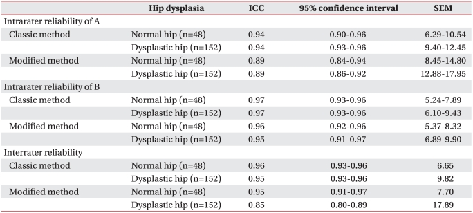
Intra-rater reliability was evaluated in both methods and groups. In the case of rater-A, ICC indicated 0.94 and 0.89 in the classic method and the modified method, respectively, irrespective of group. On the other hand, SEM indicated 8.45 to 14.80 and 12.88 to 17.95 in the normal group and the dysplasia group, respectively, and was thus lower in the normal group. In the case of rater-B, ICC in the classic method indicated 0.97 irrespective of group. In the modified method, it indicated 0.96 and 0.95 in the normal group and the dysplasia group, respectively. SEM was lower in the normal group, which was similar to that of rater-A (Table 2).
Inter-rater reliability was evaluated in both methods and in both groups. In the case of the classic method, ICC indicated 0.96 (SEM: 6.65) and 0.95 (SEM: 9.82) in the normal group and the dysplasia group respectively, and thus it was higher in the normal group. In the case of the modified method, it indicated 0.95 (SEM: 7.70) and 0.85 (SEM: 17.89) respectively. Likewise, it was higher in the normal group (Table 2).
The subluxation or dislocation of the hip joint can be conveniently identified by frontal-view radiographs. For this, various indexes including the migration index, the acetabular index and center-edge angle are widely used. The selection of such indexes should be based on the patient's age or disease. The migration index is to show how much the ossified femoral head has been dislocated from the ossified acetabular roof, which is widely used in children with cerebral palsy who have open triradiate cartilages.4,14
The integration index can be easily measured, and its SEM is within ±10%. It can be easily applied to a broad age range and thus may be the most useful index to check for subluxation of the hip joint in children with cerebral palsy.4 In many studies, follow-ups were conducted on migration indexes for several months, and as a result the significant progress of hip dysplasia was defined as where SEM was over 10%. In addition, these studies reported that the migration index was useful to evaluate the effects of therapeutic interventions, such as selective dorsal rhizotomy, hip abductors and soft tissue surgery, in the subluxation of the hip joint.15-20
A follow-up on the migration index involves the proof of a decrease or increase in hip dysplasia. In this regard, it is imperative to secure intra-rater and inter-rater reliabilities. In the study conducted by Parrott et al.5 in 2002, 20 hip joints were measured 2 times by 2 raters. Inter-rater and intra-rater reliabilities were high in that ICC was over 0.9. SEM indicated ±8.3% in relation to intra-rater reliability. This suggests that when hip migration occurred, SEM reached over 8.3% when one rater conducted a follow-up. In the study conducted by Pountney et al.6 in 2003, 40 hip joints were measured 3 times by 3 raters. SEM indicated ±7.6% and ±8.1% in relation to intra-rater reliability and inter-rater reliability respectively, which implies that the reliability was clinically reliable like previous studies.
In the study of Faraj et al.7 conducted on 44 hip joints of patients with cerebral palsy (2004), intra-rater reliability was not over 13% (95% confidence interval) but inter-rater reliability reached 21 to 23%, and thus the results were inapplicable clinically. They maintained that the migration index should be measured by one rater.
Error is caused by measurement variability, inaccurate measurements or inaccurate records. In particular, the accurate determination of the datum point is critical for measurement of the migration index.6 Nevertheless, the datum point was specified only in the study of Parrott et al.5
In this study, the datum point was specified with the classic method and the modified method, whereas intra-rater and inter-rater reliabilities showed high levels with the ICC of over 0.9, which shows that the accurate determination of the datum point raises the reliability.
In relation to intra-rater reliability, SEM was lower in the classic method than in the modified method, which was similar to the results of previous studies.
In the normal hip joint, the sourcil is evenly thick and semicircular. Its lateral part is horizontal or downwardly.9,10 In case the sourcil inclines upward or is irregular in thickness, it applies uneven stress to the hip joint. Thus, the correct form of sourcil may be a criterion for acetabular dysplasia (Fig. 1). In 2000, Kim et al.11 reported that the inter-rater error may be reduced when the lateral edge of the sourcil was used as the datum point for the measurements of the migration index and the center-edge angle. In addition, they studied the anatomic structure of the sourcil by use of MRI, 3D CT and arthrographies. According to the results, the lateral edge of the sourcil fell under the lateral center of the acetabular roof. However, on radiographs the shadow of the lateral bone meant the anterolateral edge of the acetabular roof, i.e., the inferior region of the anterior inferior iliac spine.
In previous studies, it was reported that the modified method may effectively distinguish dysplastic hip joints from normal ones.8,13 However, in this study it was found that the modified method was inferior to the classic method in intra-rater and inter-rater reliabilities. As the case stands, the modified method is likely to produce unreliable results if the migration index is measured by different raters or in cases of severe hip dysplasia.
This study was conducted to compare the classic and modified method in reliability, with children that had cerebral palsy. As mentioned, the former is to use the lateral edge of the acetabular roof as the datum point, and the latter is to use the lateral edge of the sourcil. The results showed that the lateral edge of the acetabular roof was a more effective datum point than that of the sourcil, in terms of reliability. Particularly in relation to hip dysplasia, the lateral edge of the acetabular roof was found to be more reliable as the datum point than that of the sourcil. This proves how important the datum point is in measuring the migration index of children with cerebral palsy. In conclusion, the lateral edge of the acetabular roof may be more effective than that of the sourcil.
  • 1. Gamble JG, Rinsky LA, Bleck EE. Established hip dislocation in children with cerebral palsy. Clin Orthop Relat Res 1990;253:90-99.
  • 2. Laplaza FJ, Root L. Femoral anteversion and neck-shaft angles in hip instability in cerebral palsy. J Pediatr Orthop 1994;14:719-723.
  • 3. Hagglund G, Andersson S, Duppe H, Lauge-Pedersen H, Nordmark E, Westbom L. Prevention of dislocation of the hip in children with cerebral palsy. The first ten years of a population-based prevention programme. J Bone joint Surg Br 2005;87:95-101.
  • 4. Reimers J. The stability of the hip in children. A radiological study of the results of muscle surgery in cerebral palsy. Acta Orthop Scand Suppl 1980;184:1-100.
  • 5. Parrott J, Boyd RN, Dobson F, Lancaster A, Love S, Oates J, Wolfe R, Nattrass GR, Graham HK. Hip displacement in spastic cerebral palsy: repeatability of radiologic measurement. J Pediatr Orthop 2002;22:660-667.
  • 6. Pountney T, Mandy A, Gard P. Repeatability and limits of agreement in measurement of hip migration percentage in children with bilateral cerebral palsy. Physiotherapy 2003;89:276-281.
  • 7. Faraj S, Atherton WG, Stott NS. Inter- and intra-measurer error in the measurement of Reimers' hip migration percentage. J Bone Joint Surg Br 2004;86:434-437.
  • 8. Agus H, Bicimoqlu A, Omeroglu H, Tumer Y. How should the acetabular angle of Sharp be measured on a pelvic radiograph? J Pediatr Orthop 2002;22:228-231.
  • 9. Armand M, Lepisto J, Tallroth K, Elias J, Chao E. Outcome of periacetabular osteotomy: joint contact pressure calculation using standing AP radiographs, 12 patients followed for average 2 years. Acta Orthop 2005;76:303-313.
  • 10. Broughton NS, Brougjman DI, Cole WG, Menelaus MB. Reliability of radiological measurement in the assessment of the child's hip. J Bone Joint Surg Br 1989;71:6-8.
  • 11. Kim HT, Kim JI, Yoo CI. Diagnosing childhood acetabular dysplasia using the lateral margin of the sourcil. J Pediatr Orthop 2000;20:709-717.
  • 12. Lonstein JE, Beck K. Hip dislocation and subluxation in cerebral palsy. J Pediatr Orthop 1986;6:521-526.
  • 13. Ogata S, Moriya H, Tsuchiya K, Akita T, Kamegaya N, Someya M. Acetabular cover in congenital dislocation of the hip. J Bone Joint Surg Br 1990;72:190-196.
  • 14. Scrutton D. The early management of hips in cerebral palsy. Dev Med Child Neurol 1989;31:108-116.
  • 15. Shea KG, Coleman SS, Carroll K, Stevens P, Van Boerum DH. Pemberton pericapsular osteotomy to treat a dysplastic hip in cerebral palsy. J Bone Joint Surg Am 1997;79:1342-1351.
  • 16. Cottalorda J, Gautheron V, Metton G, Charmet E, Maatougui K, Chavrier Y. Predicting the outcome of adductor tenotomy. Int Orthop 1998;22:374-379.
  • 17. Heim RC, Park TS, Vogler GP, Kaufman BA, Noetzel MJ, Ortman MR. Changes in hip migration after selective dorsal rhizotomy for spastic quadriplegia in cerebral palsy. J Neurosurg 1995;82:567-571.
  • 18. Onimus M, Allamel G, Manzone P, Laurain JM. Prevention of hip dislocation in cerebral palsy by early psoas and adductors tenotomies. J Pediatr Orthop 1991;11:432-435.
  • 19. Park TS, Vogler GP, Phillips LH, Kaufman BA, Ortman MR, McClure SM, Gaffney PE. Effects of selective dorsal rhizotomy for spastic diplegia on hip migration in cerebral palsy. Pediatr Neurosurg 1994;20:43-49.
  • 20. Reimers J, Poulsen S. Adductor transfer versus tenotomy for stability of the hip in spastic cerebral palsy. J Pediatr Orthop 1984;4:52-54.
Fig. 1
Sourcil (arrow) in the normal hip (A) and dysplastic hip (B).
arm-36-33-g001.jpg
Fig. 2
Migration Index (MI) is measured with the classic method (A) and the modified method (B). H: Hilgenreiner's line, P: Perkin's line, MI=a/b×100 (%).
arm-36-33-g002.jpg
Table 1
Intra-rater and Inter-rater Reliability of Migration Index Measured with Classic and Modified Method

ICC: Intraclass corelation coefficient, SEM: Standard error of measurement

arm-36-33-i001.jpg
Table 2
Intra-rater and Inter-rater Reliability of Migration Index According to Hip Dysplasia

ICC: Intraclass corelation coefficient, SEM: Standard error of measurement

arm-36-33-i002.jpg

Figure & Data

References

    Citations

    Citations to this article as recorded by  
    • A fully automated measurement of migration percentage on ultrasound images in children with cerebral palsy
      Reza Yousefvand, Thanh-Tu Pham, Lawrence H. Le, John Andersen, Edmond Lou
      Medical & Biological Engineering & Computing.2025; 63(4): 1177.     CrossRef
    • Botulinum toxin injections for the treatment of hip instability in the pediatric population with cerebral palsy: a systematic review
      Debra A. Sala, Eduardo del Rosario
      International Journal of Rehabilitation Research.2025; 48(1): 7.     CrossRef
    • Machine learning-assisted classification of hip conditions in pediatric cerebral palsy patients using migration percentage measurements
      Sema Ertan Birsel, Ekrem Demirci, Ali Seker, Kadriye Yasemin Usta Ayanoğlu, Emir Oncu, Fatih Ciftci
      Bone Reports.2025; 25: 101852.     CrossRef
    • Fully automated measurement of paediatric cerebral palsy pelvic radiographs with BoneFinder
      Katie Hughes, Jessenka Luzar, Jonathan Lang, Daniel C. Perry, Mark S. Gaston
      The Bone & Joint Journal.2025; 107-B(7): 752.     CrossRef
    • Radiological Assessment of Inter- and Intra-observer Reliability in Hip Migration Measurements in Children With Cerebral Palsy at a Tertiary Referral Center
      Joseph Sajeev, Binu Kurian, Jaya V Lal, Arun George, Joe Joseph
      Cureus.2025;[Epub]     CrossRef
    • Beyond words: embracing migration percentage as the universal measurement for hip displacement in children with cerebral palsy by radiologists and orthopedic surgeons
      Daniel Raftis, Sarah Dance, Laura Mazudie Ndjonko, Ahmed Elabd, Sean Tabaie
      Journal of Pediatric Orthopaedics B.2024; 33(5): 515.     CrossRef
    • Reliability of assessing proximal femur geometry with Rutz classification schema in patients with cerebral palsy
      Maciej Kasprzyk, Aleksander Koch, Lukasz M. Karbowski, Marek Jóźwiak, Unni G. Narayanan
      Journal of Pediatric Orthopaedics B.2023; 32(3): 241.     CrossRef
    • Measuring hip migration percentage in cerebral palsy using the HipScreen app
      John Amen, Oliver Perkins, Jill Cadwgan, Stephen J. Cooke, Konstantinos Kafchitsas, Michail Kokkinakis
      Bone & Joint Open.2023; 4(5): 363.     CrossRef
    • Hip reconstruction in cerebral palsy: Lessons from a single center and 137 hips
      Anoushka Ayub, Christy Graff, Lajos Maurovich Horvat, Claudia Maizen
      Journal of Children's Orthopaedics.2023; 17(5): 469.     CrossRef
    • Beyond Words: Embracing Migration Percentage as the Universal Measurement for Hip Displacement in Children With Cerebral Palsy by Radiologists and Orthopedic Surgeons
      Daniel Raftis, Sarah Dance, Laura Mazudie Ndjonko, Ahmed Elabd, Sean Tabaie
      Cureus.2023;[Epub]     CrossRef
    • Effect of different durations of using a standing frame on the rate of hip migration in children with moderate to severe cerebral palsy: a feasibility study for a randomised controlled trial
      R. Rapson, T. King, C. Morris, R. Jeffery, J. Mellhuish, C. Stephens, J. Marsden
      Physiotherapy.2022; 116: 42.     CrossRef
    • Short Term Radiological Outcome of Combined Femoral and Ilium Osteotomy in Pelvic Reconstruction of the Child
      Lorenz Pisecky, Gerhard Großbötzl, Stella Stevoska, Matthias Christoph Michael Klotz, Christina Haas, Tobias Gotterbarm, Matthias Luger, Manuel Gahleitner
      Children.2022; 9(3): 441.     CrossRef
    • A Review of Radiology Reports From Hip Surveillance Radiographs for Children With Cerebral Palsy
      Stacey D. Miller, Jaimy Coates, Jeffrey N. Bone, Jennifer Farr, Kishore Mulpuri
      Journal of Pediatric Orthopaedics.2022; 42(7): e742.     CrossRef
    • Assessment of hip displacement in children with cerebral palsy using machine learning approach
      Thanh-Tu Pham, Minh-Binh Le, Lawrence H. Le, John Andersen, Edmond Lou
      Medical & Biological Engineering & Computing.2021; 59(9): 1877.     CrossRef
    • Effect of the Sharrard procedure on hip instability in children with down syndrome: A retrospective study
      Frederike E.C.M. Mulder, Levinus A. Bok, Florens Q.M.P. van Douveren, Hans E.H. Pruijs, Adelgunde V.C.M. Zeegers
      Journal of Children's Orthopaedics.2021; 15(5): 488.     CrossRef
    • Practice Essentials of Imaging in Early Diagnosis of DDH
      Alka Karnik, Ashwin Lawande, Malini Ashwin Lawande, Deepak Patkar, Alaric Aroojis, Nidhi Bhatnagar
      Indian Journal of Orthopaedics.2021; 55(6): 1466.     CrossRef
    • Progressive lateralization and constant hip geometry in children with DDH, NDH, and LCPD following hip reconstructive surgery: a cohort study of 73 patients with a mean follow-up of 4.9 years
      Lorenz Pisecky, Gerhard Großbötzl, Manuel Gahleitner, Stella Stevoska, Christian Stadler, Christina Haas, Tobias Gotterbarm, Matthias C. Klotz
      Archives of Orthopaedic and Trauma Surgery.2021; 143(3): 1193.     CrossRef
    • Is it safe for extended-role radiographers to measure migration percentage in children with cerebral palsy?
      B.A. Marson, B.J. Oakley, S. Srinivasan, S. S, J. Chell, K. Halliday, J. Hunter, K. Price
      Radiography.2020; 26(4): e246.     CrossRef
    • 3D ultrasound to quantify lateral hip displacement in children with cerebral palsy: a validation study
      Rebecca H Kay, Jonathan J Noble, Liam Johnston, Stephen F Keevil, Michail Kokkinakis, Daniel Reed, Martin Gough, Adam P Shortland
      Developmental Medicine & Child Neurology.2020; 62(12): 1389.     CrossRef
    • Are Seating Systems With a Medial Knee Support Really Helpful for Hip Displacement in Children With Spastic Cerebral Palsy GMFCS IV and V?
      In Soo Kim, Donghwi Park, Jin Young Ko, Ju Seok Ryu
      Archives of Physical Medicine and Rehabilitation.2019; 100(2): 247.     CrossRef
    • Evaluation of errors in measurements of infantile hip radiograph using digitally reconstructed radiograph from three-dimensional MRI
      Daisuke Hamano, Kiyoshi Yoshida, Chikahisa Higuchi, Dai Otsuki, Hideki Yoshikawa, Kazuomi Sugamoto
      Journal of Orthopaedics.2019; 16(3): 302.     CrossRef
    • The migration percentage measured on EOS® standing full-leg radiographs: equivalent and advantageous in ambulant children with cerebral palsy
      Jef Neirynck, Renee Proost, Anja Van Campenhout
      BMC Musculoskeletal Disorders.2019;[Epub]     CrossRef
    • Migration percentage and odds of recurrence/subsequent surgery after treatment for hip subluxation in pediatric cerebral palsy: A meta-analysis and systematic review
      K. N. Agarwal, C. Chen, D. M. Scher, E. R. Dodwell
      Journal of Children's Orthopaedics.2019; 13(6): 582.     CrossRef
    • Femoral and Dega osteotomies in the treatment of habitual hip dislocation in down syndrome patients - is it efficient or not?
      Ahmad S. Aly, Mohamed A. Al-kersh
      Journal of Children's Orthopaedics.2018; 12(3): 227.     CrossRef
    • Treatment of the Dislocated Hip in Infants With Spasticity
      Christian A. Refakis, Keith D. Baldwin, David A. Spiegel, Wudbhav N. Sankar
      Journal of Pediatric Orthopaedics.2018; 38(7): 345.     CrossRef
    • The relationship between the level of lesion and progression in Reimer’s index of spina bifida patients
      Huseyin Gunay, Murat Celal Sozbilen, Mahmut Altınisik, Ismail Eralp Kacmaz, Elcil Kaya Bicer
      Child's Nervous System.2017; 33(2): 307.     CrossRef
    • The Effects of a Home-Based Connective Tissue Targeting Therapy on Hip Development in Children With Cerebral Palsy: Six Case Reports
      Erika Drewes, Mark Driscoll, Leonid Blyum, Diane Vincentz
      EXPLORE.2016; 12(4): 268.     CrossRef
    • Hip-joint congruity after Dega osteotomy in patients with cerebral palsy: long-term results
      Frank Braatz, Daniel Staude, Matthias C. Klotz, Sebastian I. Wolf, Thomas Dreher, Stefan Lakemeier
      International Orthopaedics.2016; 40(8): 1663.     CrossRef
    • The effect of physician experience on the measurement reliability of the Reimers’ hip migration percentage in children with cerebral palsy
      Pınar Doruk Analan, Emine Ece Yilmaz, Mehmet Adam, Berrin Leblebici
      Journal of Physical Therapy Science.2015; 27(10): 3255.     CrossRef
    • Hip problems in cerebral palsy
      Cemil Yildiz, Ismail Demirkale
      Current Opinion in Pediatrics.2014; 26(1): 85.     CrossRef
    • Hip reconstruction surgery is successful in restoring joint congruity in patients with cerebral palsy: long-term outcome
      Frank Braatz, Annette Eidemüller, Matthias C. Klotz, Nicholas A. Beckmann, Sebastian I. Wolf, Thomas Dreher
      International Orthopaedics.2014; 38(11): 2237.     CrossRef
    • Proximal femoral resection without post-operative traction for the painful dislocated hip in young patients with cerebral palsy
      J. Dartnell, M. Gough, J. M. H. Paterson, F. Norman-Taylor
      The Bone & Joint Journal.2014; 96-B(5): 701.     CrossRef
    • Validity and reliability of radiological methods to assess proximal hip geometry in children with cerebral palsy: a systematic review
      Christelle Pons, Olivier Rémy‐Néris, Béatrice Médée, Sylvain Brochard
      Developmental Medicine & Child Neurology.2013; 55(12): 1089.     CrossRef

    Download Citation

    Download a citation file in RIS format that can be imported by all major citation management software, including EndNote, ProCite, RefWorks, and Reference Manager.

    Format:

    Include:

    Reliability of Hip Migration Index in Children with Cerebral Palsy: The Classic and Modified Methods
    Ann Rehabil Med. 2012;36(1):33-38.   Published online February 29, 2012
    Download Citation
    Download a citation file in RIS format that can be imported by all major citation management software, including EndNote, ProCite, RefWorks, and Reference Manager.

    Format:
    • RIS — For EndNote, ProCite, RefWorks, and most other reference management software
    • BibTeX — For JabRef, BibDesk, and other BibTeX-specific software
    Include:
    • Citation for the content below
    Reliability of Hip Migration Index in Children with Cerebral Palsy: The Classic and Modified Methods
    Ann Rehabil Med. 2012;36(1):33-38.   Published online February 29, 2012
    Close

    Figure

    • 0
    • 1
    Reliability of Hip Migration Index in Children with Cerebral Palsy: The Classic and Modified Methods
    Image Image
    Fig. 1 Sourcil (arrow) in the normal hip (A) and dysplastic hip (B).
    Fig. 2 Migration Index (MI) is measured with the classic method (A) and the modified method (B). H: Hilgenreiner's line, P: Perkin's line, MI=a/b×100 (%).
    Reliability of Hip Migration Index in Children with Cerebral Palsy: The Classic and Modified Methods

    Intra-rater and Inter-rater Reliability of Migration Index Measured with Classic and Modified Method

    ICC: Intraclass corelation coefficient, SEM: Standard error of measurement

    Intra-rater and Inter-rater Reliability of Migration Index According to Hip Dysplasia

    ICC: Intraclass corelation coefficient, SEM: Standard error of measurement

    Table 1 Intra-rater and Inter-rater Reliability of Migration Index Measured with Classic and Modified Method

    ICC: Intraclass corelation coefficient, SEM: Standard error of measurement

    Table 2 Intra-rater and Inter-rater Reliability of Migration Index According to Hip Dysplasia

    ICC: Intraclass corelation coefficient, SEM: Standard error of measurement

    TOP