
• KARM
  • Contact us
  • E-Submission
ABOUT
ARTICLE TYPES
BROWSE ARTICLES
AUTHOR INFORMATION

Page Path

8
results for

"Tractography"

Filter

Article category

Publication year

"Tractography"

Original Articles

Spinal cord injury

Quantitative Analysis in Cervical Spinal Cord Injury Patients Using Diffusion Tensor Imaging and Tractography
Geun Seok Park, Tae Uk Kim, Seong Jae Lee, Jung Keun Hyun, Seo Young Kim
Ann Rehabil Med 2022;46(4):172-184.   Published online August 31, 2022
DOI: https://doi.org/10.5535/arm.22053
Objective
To investigate the clinical usefulness of diffusion tensor imaging (DTI) and tractography in the prediction of outcomes after traumatic cervical spinal cord injury (SCI) and to assess whether the predictability is different between DTI and tractography administered before and after surgery.
Methods
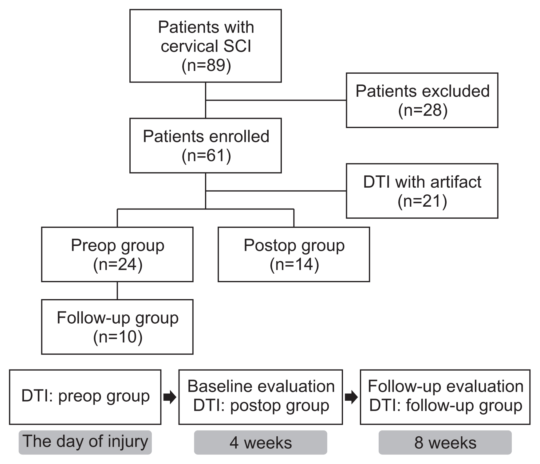
Sixty-one subjects with traumatic cervical SCI were randomly assigned to preop or postop groups and received DTI accordingly. Among the patients who had DTI before surgery, we assigned 10 patients who had received repeated DTI examinations at 8 weeks after injury to the follow-up group. Fractional anisotropy (FA) and apparent diffusion coefficient (ADC) values were obtained from DTI, and imaginary fiber and crossing fiber numbers were calculated from the tractography. Neurological status and functional status were assessed at 4 and 8 weeks after SCI.
Results
The neurologic and functional statuses of both groups improved after 4 weeks. Out of the initial 61 patients who were enrolled in the study, the failure rate of DTI image analysis was significantly higher in the postop group (n=17, 41.5%) than in the preop group (n=6, 20%). The FA values and fiber numbers in the preop group tended to be higher than those in the postop group, whereas ADC values were lower in the preop group. When comparing the tractographic findings in the follow-up group, imaginary fiber numbers at the C6 and C7 levels and crossing fiber numbers from the C3 to C6 levels were significantly decreased after surgery. Several DTI and tractographic parameters (especially the ADC value at the C4 level and imaginary fiber numbers at the C6 level) showed significant correlations with neurologic and functional statuses in both the preop and postop groups. These findings were most prominent when DTI and physical examination were simultaneously performed.
Conclusion
Preoperative DTI and tractography demonstrated better FA and ADC values with lower interpretation failure rates than those obtained after surgery, whereas postoperative data significantly reflected the patient’s clinical state at the time of evaluation. Therefore, DTI and tractography could be useful in predicting clinical outcomes after traumatic cervical SCI and should be interpreted separately before and after spine surgery.

Citations

Citations to this article as recorded by  
  • Diffusion tractography outside the brain: the road less travelled
    Kurt G. Schilling, Irvin Teh, Julien Cohen-Adad, Richard Dortch, Ibrahim Ibrahim, Nian Wang, Bruce Damon, Rory L. Cochran, Alexander Leemans
    Brain Structure and Function.2026;[Epub]     CrossRef
  • Intravenous Infusion of Mesenchymal Stem Cells Enhances Long-Tract Reorganization That Circumvents the Lesion in a Rat Staggered Hemisection Spinal Cord Injury Model
    Hisashi Obara, Masahito Nakazaki, Takahiro Yokoyama, Hiroshi Nagahama, Ryunosuke Fukushi, Shinichi Oka, Ryo Ukai, Yuko Kataoka-Sasaki, Masanori Sasaki, Jeffery D. Kocsis, Atsushi Teramoto, Toshihiko Yamashita, Osamu Honmou
    Biomedicines.2026; 14(9): 1979.     CrossRef
  • Negative Correlation Between Fractional Anisotropy on Diffusion Tensor Imaging and Neck Disability Index in Cervical Spondylotic Radiculopathy After Percutaneous Cervical Nucleoplasty: A Cross-Sectional Study
    Ning Fan, Tusheng Li, Xuanyu Lu, Xinyu Ma, Lihui Yang, Peng Du, Lei Zang, Shuo Yuan
    Journal of Pain Research.2025; Volume 18: 3977.     CrossRef
  • Quantitative Magnetic Resonance Identifies Recovery from Spinal Cord Injury after Bioactive Implants
    DIANA OSORIO-LONDOÑO, AXAYÁCATL MORALES-GUADARRAMA, ROBERTO OLAYO-GONZÁLEZ, ERNESTO ROLDAN-VALADEZ
    Archives of Medical Research.2024; 55(5): 103012.     CrossRef
  • 9,127 View
  • 143 Download
  • 3 Web of Science
  • 4 Crossref
Objective

To correlate the resting metabolism of hand knob and hand function after stroke, diffuse tensor tractography (DTT) and 18F-fluorodeoxyglucose position emission tomography (18F-FDG PET) were used to evaluate constructible state of white matter tract and metabolic state of gray matter, respectively.

Methods

A total of 17 patients were included in the study, who had suffered a stroke with hand weakness, after a stroke. They underwent diffusion tensor analysis and FDG PET in the subacute period. The ratio of both hemisphere parameters in voxel number of fibers, fractional anisotropy (FA) and apparent diffusion coefficient obtained by corticospinal tract as constructed by DTT, and the metabolism of hand knob area on cerebral cortex obtained from 18F-FDG PET were calculated. Hand movement scale was evaluated on the day of FDG PET or tractography, and at 6 months after onset.

Results

Difference of FA in DTT between both hemispheres and hand knob metabolism in FDG PET significantly correlated with the hand movement scale at the subacute stage and 6 months after onset. However, the difference of both hemispheres in DTT and metabolism of hand knob area was not significant.

Conclusion

Resting metabolism on hand knob in FDG PET correlated with hand function after stroke.

Citations

Citations to this article as recorded by  
  • A Narrative Review of Stroke of Cortical Hand Knob Area
    Jamir Pitton Rissardo, Vishnu Vardhan Byroju, Sushni Mukkamalla, Ana Letícia Fornari Caprara
    Medicina.2024; 60(2): 318.     CrossRef
  • Association between human gray matter metabotropic glutamate receptor-5 availability in vivo and white matter properties: a [11C]ABP688 PET and diffusion tensor imaging study
    Song E. Kim, Yo-Han Joo, Young-Don Son, Hang-Keun Kim, Sang-Yoon Lee, Min-Woo Lee, Tatsuo Ido, Jong-Hoon Kim
    Brain Structure and Function.2020; 225(6): 1805.     CrossRef
  • The reliability of identifying the Omega sign using axial T2-weighted magnetic resonance imaging
    Hesham Mostafa Zakaria, Peter Joseph Massa, Richard L Smith, Tarek Hazem Moharram, John Corrigan, Ian Lee, Lonni Schultz, Jianhui Hu, Suresh Patel, Brent Griffith
    The Neuroradiology Journal.2018; 31(4): 345.     CrossRef
  • 8,150 View
  • 57 Download
  • 3 Web of Science
  • 3 Crossref
Comparison of Diffusion Tensor Tractography and Motor Evoked Potentials for the Estimation of Clinical Status in Subacute Stroke
Kwang-Soo Chun, Yong-Taek Lee, Jong-Wan Park, Joon-Youn Lee, Chul-Hyun Park, Kyung Jae Yoon
Ann Rehabil Med 2016;40(1):126-134.   Published online February 26, 2016
DOI: https://doi.org/10.5535/arm.2016.40.1.126
Objective

To compare diffusion tensor tractography (DTT) and motor evoked potentials (MEPs) for estimation of clinical status in patients in the subacute stage of stroke.

Methods

Patients with hemiplegia due to stroke who were evaluated using both DTT and MEPs between May 2012 and April 2015 were recruited. Clinical assessments investigated upper extremity motor and functional status. Motor status was evaluated using Medical Research Council grading and the Fugl-Meyer Assessment of upper limb and hand (FMA-U and FMA-H). Functional status was measured using the Modified Barthel Index (MBI). Patients were classified into subgroups according to DTT findings, MEP presence, fractional anisotropy (FA) value, FA ratio (rFA), and central motor conduction time (CMCT). Correlations of clinical assessments with DTT parameters and MEPs were estimated.

Results

Fifty-five patients with hemiplegia were recruited. In motor assessments (FMA-U), MEPs had the highest sensitivity and negative predictive value (NPV) as well as the second highest specificity and positive predictive value (PPV). CMCT showed the highest specificity and PPV. Regarding functional status (MBI), FA showed the highest sensitivity and NPV, whereas CMCT had the highest specificity and PPV. Correlation analysis showed that the resting motor threshold (RMT) ratio was strongly associated with motor status of the upper limb, and MEP parameters were not associated with MBI.

Conclusion

DTT and MEPs could be suitable complementary modalities for analyzing the motor and functional status of patients in the subacute stage of stroke. The RMT ratio was strongly correlated with motor status.

Citations

Citations to this article as recorded by  
  • Short-term Efficacy of Hand-Arm Bimanual Intensive Training on Upper Arm Function in Acute Stroke Patients: A Randomized Controlled Trial
    Guilin Meng, Xiuling Meng, Yan Tan, Jia Yu, Aiping Jin, Yanxin Zhao, Xueyuan Liu
    Frontiers in Neurology.2018;[Epub]     CrossRef
  • The role of diffusion tensor imaging as an objective tool for the assessment of motor function recovery after paraplegia in a naturally-occurring large animal model of spinal cord injury
    Adriano Wang-Leandro, Marc K. Hobert, Sabine Kramer, Karl Rohn, Veronika M. Stein, Andrea Tipold
    Journal of Translational Medicine.2018;[Epub]     CrossRef
  • Spontaneous acute and chronic spinal cord injuries in paraplegic dogs: a comparative study of in vivo diffusion tensor imaging
    A Wang-Leandro, M K Hobert, N Alisauskaite, P Dziallas, K Rohn, V M Stein, A Tipold
    Spinal Cord.2017; 55(12): 1108.     CrossRef
  • 7,365 View
  • 58 Download
  • 3 Web of Science
  • 3 Crossref
Prediction of Motor Recovery Using Diffusion Tensor Tractography in Supratentorial Stroke Patients With Severe Motor Involvement
Kang Hee Kim, Yun-Hee Kim, Min Su Kim, Chang-hyun Park, Ahee Lee, Won Hyuk Chang
Ann Rehabil Med 2015;39(4):570-576.   Published online August 25, 2015
DOI: https://doi.org/10.5535/arm.2015.39.4.570
Objective

To investigate whether early stage diffusion tensor tractography (DTT) values predict motor function at 3 months after onset in supratentorial stroke patients with severe motor involvement.

Methods

A retrospective study design was used to analyze medical records and neuroimaging data of 49 supratentorial stroke patients with severe motor involvement. Diffusion tensor imaging was assessed within 3 weeks after stroke in all patients. Three-dimensional tractography of the ipsilateral corticospinal tract (CST) was performed using the fiber assignment of the continuous tracking algorithm. The two-step DTT analysis was used. The first step was classification according to ipsilateral CST visualization. The second step was a quantitative analysis of the visible-CST group parameters. Motor function was assessed at 2 weeks and at 3 months after stroke. Comparative and correlation analyses were performed between DTT-derived measures and motor assessment scores.

Results

Motor function of the upper extremity at 3 months after stroke was significantly higher in the visible-CST group than that in the nonvisible-CST group (p<0.05). Early stage fractional anisotropy was of DTT correlated significantly with upper extremity motor function at 3 months after stroke in the visible-CST group (p<0.05).

Conclusion

These results demonstrate that early DTT-derived measures predict motor recovery in the upper extremity at 3 months after onset in supratentorial stroke patients with severe motor involvement.

Citations

Citations to this article as recorded by  
  • The relationship between corticospinal tract and the affected lower limb function and walking ability in subacute stroke patients: a preliminary study
    Tomohiro Oyamada, Masaki Kobayashi, Kentaro Yotsumoto, Takeshi Kawarabayashi, Shigeru Usuda
    Journal of Physical Therapy Science.2026; 38(3): 117.     CrossRef
  • Predictive value of corticospinal tract integrity and clinical factors for post-stroke motor recovery using diffusion tensor imaging
    Jihee Park, Sekwang Lee, Yoonhye Na, Minjae Cho, Sung-Bom Pyun
    Clinical Neurophysiology.2026; 186: 2111848.     CrossRef
  • Objective motor function assessment using diffusion tensor tractography in subacute acquired brain injury: A retrospective observational study
    Au-Jin Wang, Choong-Hee Roh, Myoung-Hwan Ko, Da-Sol Kim, Yu Hui Won, Sung-Hee Park, Jeong-Hwan Seo, Gi-Wook Kim
    Medicine.2025; 104(52): e46721.     CrossRef
  • Evaluating the axonal injury and predicting the motor function recovery in supratentorial acute stroke patients
    Anh Tuan Tran, Van Tuan Nguyen, Quang Huy Huynh, Dinh Minh Nguyen, Huy Manh Bui, Hai Dang Vu, Tuan Vu Nguyen, Thu Ha Nguyen-Thi
    Interdisciplinary Neurosurgery.2024; 36: 101919.     CrossRef
  • Neuroimaging of motor recovery after ischemic stroke − functional reorganization of motor network
    Pei Yu, Ruoyu Dong, Xiao Wang, Yuqi Tang, Yaning Liu, Can Wang, Ling Zhao
    NeuroImage: Clinical.2024; 43: 103636.     CrossRef
  • Mối tương quan giữa một số chỉ số trên cộng hưởng từ bó tháp trong tiên lượng khả năng phục hồi chức năng vận động ở bệnh nhân nhồi máu não vùng trên lều
    Thị Phương Lâm Trịnh, Thị Thanh Bình Nguyễn, Thị Mỹ Lê, Công Tiến Nguyễn
    Tạp chí thần kinh học Việt Nam.2024; (43): 42.     CrossRef
  • A Diffusion Tensor Imaging–Based Prognostic Classification for Surgery of Intrinsic Lesions Involving the Motor Pathways
    Giovanni Muscas, Antonio Pisano, Riccardo Carrai, Andrea Bianchi, Federico Capelli, Vita Maria Montemurro, Cristiana Martinelli, Enrico Fainardi, Antonello Grippo, Alessandro Della Puppa
    World Neurosurgery.2023; 172: e565.     CrossRef
  • Beyond Diffusion Tensor MRI Methods for Improved Characterization of the Brain after Ischemic Stroke: A Review
    E.V.R. DiBella, A. Sharma, L. Richards, V. Prabhakaran, J.J. Majersik, S.K. HashemizadehKolowri
    American Journal of Neuroradiology.2022; 43(5): 661.     CrossRef
  • Relationship between size and location of infarction beside lateral ventricle and motor recovery following rehabilitation
    You-Kui Lv, Li-Ping Huang, Zhuang-Wei Fang, Gang Wang, Li-Kang Wang, Ming Zhou, Xin-Ling Su, Dan-Yang Ding, Xing-Lin Wang
    NeuroRehabilitation.2022; 51(3): 527.     CrossRef
  • Elucidating the mechanisms of post-stroke motor recovery mediated by electroacupuncture using diffusion tensor tractography
    Min Su Kim, Byung Soon Moon, Jae-yoon Ahn, Sang-song Shim, Jong-Min Yun, Min Cheol Joo
    Frontiers in Neurology.2022;[Epub]     CrossRef
  • An overview of fractional anisotropy as a reliable quantitative measurement for the corticospinal tract (CST) integrity in correlation with a Fugl-Meyer assessment in stroke rehabilitation
    Mohd Khairul Izamil Zolkefley, Younis M. S. Firwana, Hasnettty Zuria Mohamed Hatta, Christina Rowbin, Che Mohd Nasril Che Mohd Nassir, Muhammad Hafiz Hanafi, Mohd Shafie Abdullah, Muzaimi Mustapha
    Journal of Physical Therapy Science.2021; 33(1): 75.     CrossRef
  • Proprioception and motor performance after stroke: An examination of diffusion properties in sensory and motor pathways
    Sonja E. Findlater, Erin L. Mazerolle, G. Bruce Pike, Sean P. Dukelow
    Human Brain Mapping.2019; 40(10): 2995.     CrossRef
  • Brain networks and their relevance for stroke rehabilitation
    Adrian G. Guggisberg, Philipp J. Koch, Friedhelm C. Hummel, Cathrin M. Buetefisch
    Clinical Neurophysiology.2019; 130(7): 1098.     CrossRef
  • Can the integrity of the corticospinal tract predict the long-term motor outcome in poststroke hemiplegic patients?
    Ae Ryoung Kim, Dae Hyun Kim, So Young Park, Sunghyon Kyeong, Yong Wook Kim, Seung Koo Lee, Deog Young Kim
    NeuroReport.2018; 29(6): 453.     CrossRef
  • On the validity of lesion-behaviour mapping methods
    Christoph Sperber, Hans-Otto Karnath
    Neuropsychologia.2018; 115: 17.     CrossRef
  • Prediction of lower extremity motor recovery in persons with severe lower extremity paresis after stroke
    Sheau-Ling Huang, Bang-Bin Chen, I-Ping Hsueh, Jiann-Shing Jeng, Chia-Lin Koh, Ching-Lin Hsieh
    Brain Injury.2018; 32(5): 627.     CrossRef
  • Utility of Fractional Anisotropy in Cerebral Peduncle for Stroke Outcome Prediction: Comparison of Hemorrhagic and Ischemic Strokes
    Tetsuo Koyama, Masatoshi Koumo, Yuki Uchiyama, Kazuhisa Domen
    Journal of Stroke and Cerebrovascular Diseases.2018; 27(4): 878.     CrossRef
  • Can Neurological Biomarkers of Brain Impairment Be Used to Predict Poststroke Motor Recovery? A Systematic Review
    Bokkyu Kim, Carolee Winstein
    Neurorehabilitation and Neural Repair.2017; 31(1): 3.     CrossRef
  • Longitudinal Structural and Functional Differences Between Proportional and Poor Motor Recovery After Stroke
    Adrian G. Guggisberg, Pierre Nicolo, Leonardo G. Cohen, Armin Schnider, Ethan R. Buch
    Neurorehabilitation and Neural Repair.2017; 31(12): 1029.     CrossRef
  • Contribution of Neuro-Imaging for Prediction of Functional Recovery after Ischemic Stroke
    Wolf-Dieter Heiss
    Cerebrovascular Diseases.2017; 44(5-6): 266.     CrossRef
  • Association Between Brain-Derived Neurotrophic Factor Genotype and Upper Extremity Motor Outcome After Stroke
    Won Hyuk Chang, Eunhee Park, Jungsoo Lee, Ahee Lee, Yun-Hee Kim
    Stroke.2017; 48(6): 1457.     CrossRef
  • Predicting motor improvement after stroke with clinical assessment and diffusion tensor imaging
    Ethan R. Buch, Sviatlana Rizk, Pierre Nicolo, Leonardo G. Cohen, Armin Schnider, Adrian G. Guggisberg
    Neurology.2016; 86(20): 1924.     CrossRef
  • 8,439 View
  • 88 Download
  • 19 Web of Science
  • 22 Crossref

Case Reports

Diffusion Tensor Tractography in Two Cases of Kernohan-Woltman Notch Phenomenon
Seung-Gul Jang, Sung-Bom Pyun
Ann Rehabil Med 2013;37(6):879-885.   Published online December 23, 2013
DOI: https://doi.org/10.5535/arm.2013.37.6.879

Kernohan-Woltman notch phenomenon (KWP) is an ipsilateral motor weakness due to compression of the contralateral cerebral peduncle. We report two cases of KWP following traumatic brain injury. In case 1, ipsilateral hemiplegia was noted after right subdural hemorrhage. Although magnetic resonance imaging showed no abnormal signal changes on cerebral peduncle, diffusion tensor tractography (DTT) revealed interruption of corticospinal tract (CST) at lower level of the midbrain level. In case 2, there was abnormal signal change of the right cerebral peduncle contralateral to the primary lesion and we could not reconstruct right CST. Case 1 showed unsatisfactory motor recovery even after 15 months, and follow-up DTT showed no change. In case 2, follow-up DTT was not performed, but her ipsilateral hemiparesis had almost disappeared during the 15 months. DTT would be useful in detecting ipsilateral hemiparesis due to KWP and the clinical course may differ according to the lesion characteristics.

Citations

Citations to this article as recorded by  
  • The Kernohan-Woltman notch phenomenon model of brainstem injury: Etiopathogenic classification and clinical outcome
    R. Carrasco Moro, L. Barrios, J.M. Pascual
    Revue Neurologique.2026;[Epub]     CrossRef
  • Paradoxical hemiparesis caused by acute subdural hematoma: Kernohan-Woltman notch phenomenon
    Zhongjing Zhang, Dong Xie, Pengfei Yao, Jie Zhou
    Asian Journal of Surgery.2025; 48(8): 4833.     CrossRef
  • A Discussion of a Case of Paradoxical Ipsilateral Hemiparesis in a Patient Diagnosed with Pterional Meningioma
    Ligia Gabriela Tataranu
    Journal of Clinical Medicine.2025; 14(8): 2689.     CrossRef
  • Kernohan-Woltman notch phenomenon: an exceptional neurological picture?
    R. Carrasco Moro, J.M. Pascual Garvi, C. Vior Fernández, E.E. Espinosa Rodríguez, G. Martín Palomeque, L. Cabañes Martínez, M. López Gutiérrez, A. Acitores Cancela, E. Barrero Ruiz, J.S. Martínez San Millán
    Neurología (English Edition).2024; 39(8): 683.     CrossRef
  • In Reply to the Letter to the Editor Regarding “Kernohan-Woltman Notch Phenomenon Following Acute Subdural Hematoma”
    Nathan Beucler
    World Neurosurgery.2024; 188: 244.     CrossRef
  • Fenómeno de Kernohan-Woltman: ¿un cuadro neurológico excepcional?
    R. Carrasco Moro, J.M. Pascual Garvi, C. Vior Fernández, E.E. Espinosa Rodríguez, G. Martín Palomeque, L. Cabañes Martínez, M. López Gutiérrez, A. Acitores Cancela, E. Barrero Ruiz, J.S. Martínez San Millán
    Neurología.2024; 39(8): 683.     CrossRef
  • Beyond uncal herniation: An updated diagnostic reappraisal of ipsilateral hemiparesis and the Kernohan-Woltman notch phenomenon
    R. Carrasco-Moro, J.S. Martínez-San Millán, J.M. Pascual
    Revue Neurologique.2023; 179(8): 844.     CrossRef
  • The Kernohan-Woltman Notch Phenomenon : A Systematic Review of Clinical and Radiologic Presentation, Surgical Management, and Functional Prognosis
    Nathan Beucler, Pierre-Julien Cungi, Guillaume Baucher, Stéphanie Coze, Arnaud Dagain, Pierre-Hugues Roche
    Journal of Korean Neurosurgical Society.2022; 65(5): 652.     CrossRef
  • A case of Kernohan-Woltman notch phenomenon caused by an epidural hematoma: the diagnostic and prognostic value of PET/CT imaging
    Yuliang Lin, Alan Chen-Lung Chou, Xiangming Lin, Zhende Wu, Qichao Ju, Yuexuan Li, Zulong Ye, Bo Zhang
    BMC Neurology.2022;[Epub]     CrossRef
  • In vivo demonstration of selective vulnerability of dopamine neurons
    Dallah Yoo, Hyug-Gi Kim, Ji-In Bang, Kyung Mi Lee, Tae-Beom Ahn
    Journal of the Neurological Sciences.2020; 415: 116946.     CrossRef
  • Ipsilateral hemiparesis: the forgotten history of this paradoxical neurological sign
    Rodrigo Carrasco-Moro, Ines Castro-Dufourny, Juan S. Martínez-San Millán, Lidia Cabañes-Martínez, José M. Pascual
    Neurosurgical Focus.2019; 47(3): E7.     CrossRef
  • Kernohan-Woltman Notch Phenomenon Caused by a Traumatic Epidural Hematoma
    Jin-Hyung Lee, Mi-Ri Kang, Sang Jin Kim, Bong-Goo Yoo, Eung Gyu Kim, Ki-Hwan Ji
    Journal of Neurosonology and Neuroimaging.2019; 11(2): 182.     CrossRef
  • Kernohan–Woltman notch phenomenon: a review article
    C. H. Zhang, R. M. DeSouza, J. S. B. Kho, S. Vundavalli, G. Critchley
    British Journal of Neurosurgery.2017; 31(2): 159.     CrossRef
  • 7,194 View
  • 74 Download
  • 13 Crossref
Fornix Injury in a Patient with Rotavirus Encephalopathy: Diffusion Tensor Tractography Study
Su Min Son, Sung Ho Jang, Eun Sil Lee, Sang Ho Ahn, Dong Gyu Lee, Hee Kyung Cho
Ann Rehabil Med 2012;36(4):551-555.   Published online August 27, 2012
DOI: https://doi.org/10.5535/arm.2012.36.4.551

Rotavirus encephalopathy (RE) is a benign afebrile seizure associated with acute gastroenteritis caused by rotavirus infection. We investigated the diffusion tensor tractography (DTT) findings of a patient with RE. The patient was a 30-month-old female that had experienced a brief, generalized convulsive seizure. On the day of admission, the patient had vomiting and experienced watery diarrhea. Her stool was positive for rotavirus antigen. At onset, the patient displayed a drowsy and delirious mental status; later, a splenial lesion of the corpus callosum was found on MRI. One week later, the patient's condition improved and the splenial lesion had disappeared by conventional MRI. Initial DTI showed decreased fractional anisotropy (FA) values of fornix, as well as of the corpus callosum. A follow-up DTT showed a restored interrupted right fonical crus and increased FA values of corpus callosum and fornix. These results highlight the implications of the probability of not only a corpus callosum injury, but a fornix injury as well, in this patient with RE.

Citations

Citations to this article as recorded by  
  • Neurological complications of rotavirus infection in children: A systematic review and meta‐analysis
    A. Meyer, C. Mazzara, S. A. G. Lava, G. Treglia, M. G. Bianchetti, B. Goeggel Simonetti, G. D. Simonetti
    Acta Paediatrica.2023; 112(7): 1565.     CrossRef
  • The Fornix: Functional Anatomy, Normal Neuroimaging, and Various Pathological Conditions
    Young Jae Choi, Eun Ja Lee, Jung Eun Lee
    Investigative Magnetic Resonance Imaging.2021; 25(2): 59.     CrossRef
  • Lesions of the Corpus Callosum and Other Commissural Fibers: Diffusion Tensor Studies
    Christopher G. Filippi, Keith A. Cauley
    Seminars in Ultrasound, CT and MRI.2014; 35(5): 445.     CrossRef
  • 6,033 View
  • 27 Download
  • 3 Crossref
Original Articles
Transcallosal Fibers from Corticospinal Tract in Patients with Cerebral Hemorrage.
Kim, Wook Ro , Chang, Min Cheol , Park, Kyung A , Kwak, So Young , Do, Sung Jin , Jang, Sung Ho , Ahn, Sang Ho , Son, Su Min
J Korean Acad Rehabil Med 2009;33(6):657-663.
Objective
To investigate the relationship between the transcallosal fibers (TCFs) from the corticospinal tract (CST) and the motor function of the affected extremities in patients with cerebral hemorrhage, using diffusion tensor image tractography (DTT). Method: 49 patients with cerebral hemorrhage and 38 controls were recruited. DTT was performed using 1.5 T magnetic resonance imaging. The DTT findings were classified into 3 groups according to targeting location: no TCF from the CST (type A), TCF ended in the corpus callosum or connected to the cortex of the opposite hemisphere (type B), and TCF that descending toward the lesion after passing through the corpus callosum (type C). Results: The TCF originated from the CST of the unaffected hemisphere, and the presence of fiber descending toward the lesion after passing through the corpus callosum were significantly more prevalent in the patients who showed the worse motor function. Conclusion: The TCF originated from the unaffected CST toward the lesion in patients with cerebral hemorrhage might play a relevant role in compensating motor deficits in the major corticospinal disruption. (J Korean Acad Rehab Med 2009; 33: 657-663)
  • 2,297 View
  • 10 Download
Effect of Premotor Cortex Stimulation on Motor Learning in Basal Ganglial Hemorrhage Patients.
Kang, Ga Young , Jung, Kwang Ik , Ohn, Suk Hoon , Yoo, Woo Kyoung
J Korean Acad Rehabil Med 2011;35(2):180-187.
Objective
To investigate the effects of high frequency (10 Hz) repetitive transcranial magnetic stimulation (rTMS) on visuospatial motor learning, stimulated on the premotor cortex in basal ganglia hemorrhage patients. Method Nine patients were randomized to receive real and sham rTMS. We subdivided into two groups according to the integrity of the corticospinal tract measured by diffusion tensor tractography. The implicit visuospatial learning paradigm composed of numbers 1 to 4, in which 12 sequential numbers (2-3-1-4-3-2-4-1-3-4-2-1) were incorporated randomly. We obtained the mean fraction anisotrophy (FA) and apparent diffusion coefficient (ADC) values from the corticospinal tract and subdividing into two groups by calculating the relative value (laterality %). Results rTMS on the premotor cortex was effective only in simple motor learning but not in visuospatial learning in group analysis. The primary motor cortex excitability after the premotor cortex stimulation has been changed significantly. Subdividing into two groups according to the integrity of the corticospinal tract using the ADC value, the low ADC value group showed significant reduction of the visuospatial response time. Conclusion High frequency rTMS on the premotor cortex was effective in simple motor learning and also in the group who maintained more integrity of the corticospinal tract in basal ganglia hemorrhage.
  • 1,948 View
  • 11 Download
TOP