Objective To investigate the clinical usefulness of diffusion tensor imaging (DTI) and tractography in the prediction of outcomes after traumatic cervical spinal cord injury (SCI) and to assess whether the predictability is different between DTI and tractography administered before and after surgery.
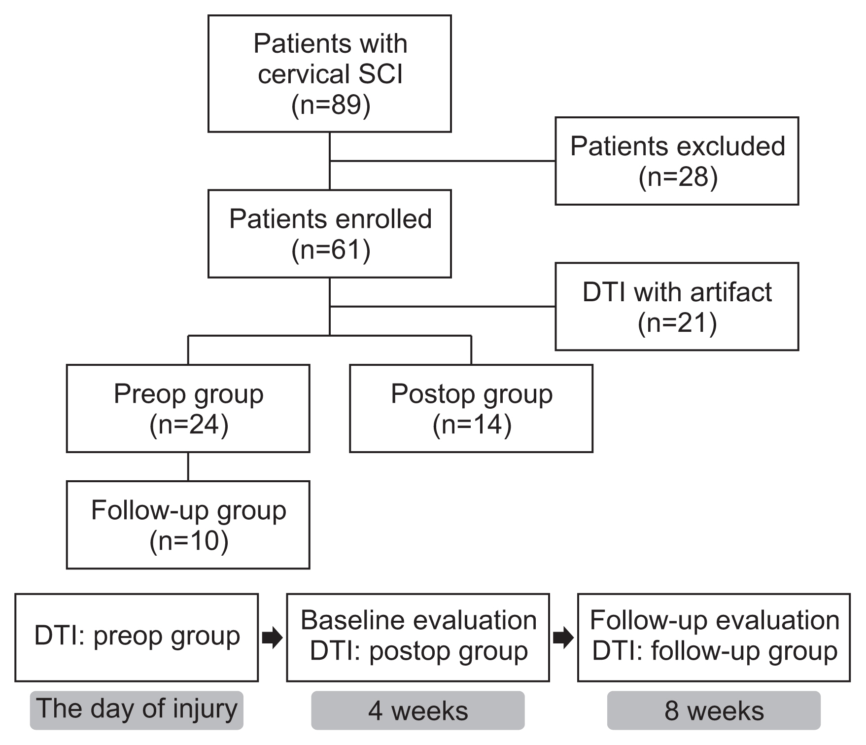
Methods Sixty-one subjects with traumatic cervical SCI were randomly assigned to preop or postop groups and received DTI accordingly. Among the patients who had DTI before surgery, we assigned 10 patients who had received repeated DTI examinations at 8 weeks after injury to the follow-up group. Fractional anisotropy (FA) and apparent diffusion coefficient (ADC) values were obtained from DTI, and imaginary fiber and crossing fiber numbers were calculated from the tractography. Neurological status and functional status were assessed at 4 and 8 weeks after SCI.
Results The neurologic and functional statuses of both groups improved after 4 weeks. Out of the initial 61 patients who were enrolled in the study, the failure rate of DTI image analysis was significantly higher in the postop group (n=17, 41.5%) than in the preop group (n=6, 20%). The FA values and fiber numbers in the preop group tended to be higher than those in the postop group, whereas ADC values were lower in the preop group. When comparing the tractographic findings in the follow-up group, imaginary fiber numbers at the C6 and C7 levels and crossing fiber numbers from the C3 to C6 levels were significantly decreased after surgery. Several DTI and tractographic parameters (especially the ADC value at the C4 level and imaginary fiber numbers at the C6 level) showed significant correlations with neurologic and functional statuses in both the preop and postop groups. These findings were most prominent when DTI and physical examination were simultaneously performed.
Conclusion Preoperative DTI and tractography demonstrated better FA and ADC values with lower interpretation failure rates than those obtained after surgery, whereas postoperative data significantly reflected the patient’s clinical state at the time of evaluation. Therefore, DTI and tractography could be useful in predicting clinical outcomes after traumatic cervical SCI and should be interpreted separately before and after spine surgery.
Citations
Citations to this article as recorded by
Diffusion tractography outside the brain: the road less travelled Kurt G. Schilling, Irvin Teh, Julien Cohen-Adad, Richard Dortch, Ibrahim Ibrahim, Nian Wang, Bruce Damon, Rory L. Cochran, Alexander Leemans Brain Structure and Function.2026;[Epub] CrossRef
Intravenous Infusion of Mesenchymal Stem Cells Enhances Long-Tract Reorganization That Circumvents the Lesion in a Rat Staggered Hemisection Spinal Cord Injury Model Hisashi Obara, Masahito Nakazaki, Takahiro Yokoyama, Hiroshi Nagahama, Ryunosuke Fukushi, Shinichi Oka, Ryo Ukai, Yuko Kataoka-Sasaki, Masanori Sasaki, Jeffery D. Kocsis, Atsushi Teramoto, Toshihiko Yamashita, Osamu Honmou Biomedicines.2026; 14(9): 1979. CrossRef
Negative Correlation Between Fractional Anisotropy on Diffusion Tensor Imaging and Neck Disability Index in Cervical Spondylotic Radiculopathy After Percutaneous Cervical Nucleoplasty: A Cross-Sectional Study Ning Fan, Tusheng Li, Xuanyu Lu, Xinyu Ma, Lihui Yang, Peng Du, Lei Zang, Shuo Yuan Journal of Pain Research.2025; Volume 18: 3977. CrossRef
Quantitative Magnetic Resonance Identifies Recovery from Spinal Cord Injury after Bioactive Implants DIANA OSORIO-LONDOÑO, AXAYÁCATL MORALES-GUADARRAMA, ROBERTO OLAYO-GONZÁLEZ, ERNESTO ROLDAN-VALADEZ Archives of Medical Research.2024; 55(5): 103012. CrossRef



 First
 Prev


